Radiographs of the brain
Lateral pneumoencephalogram
Stanford holds the copyright to the David L. Bassett anatomical images and has assigned
Creative Commons license Attribution-Share
Alike 4.0 International to all of the images.
For additional information regarding use and permissions,
please contact Dr. Drew Bourn at dbourn@stanford.edu.
Image #34-4
Radiographs of the brain
Lateral pneumoencephalogram
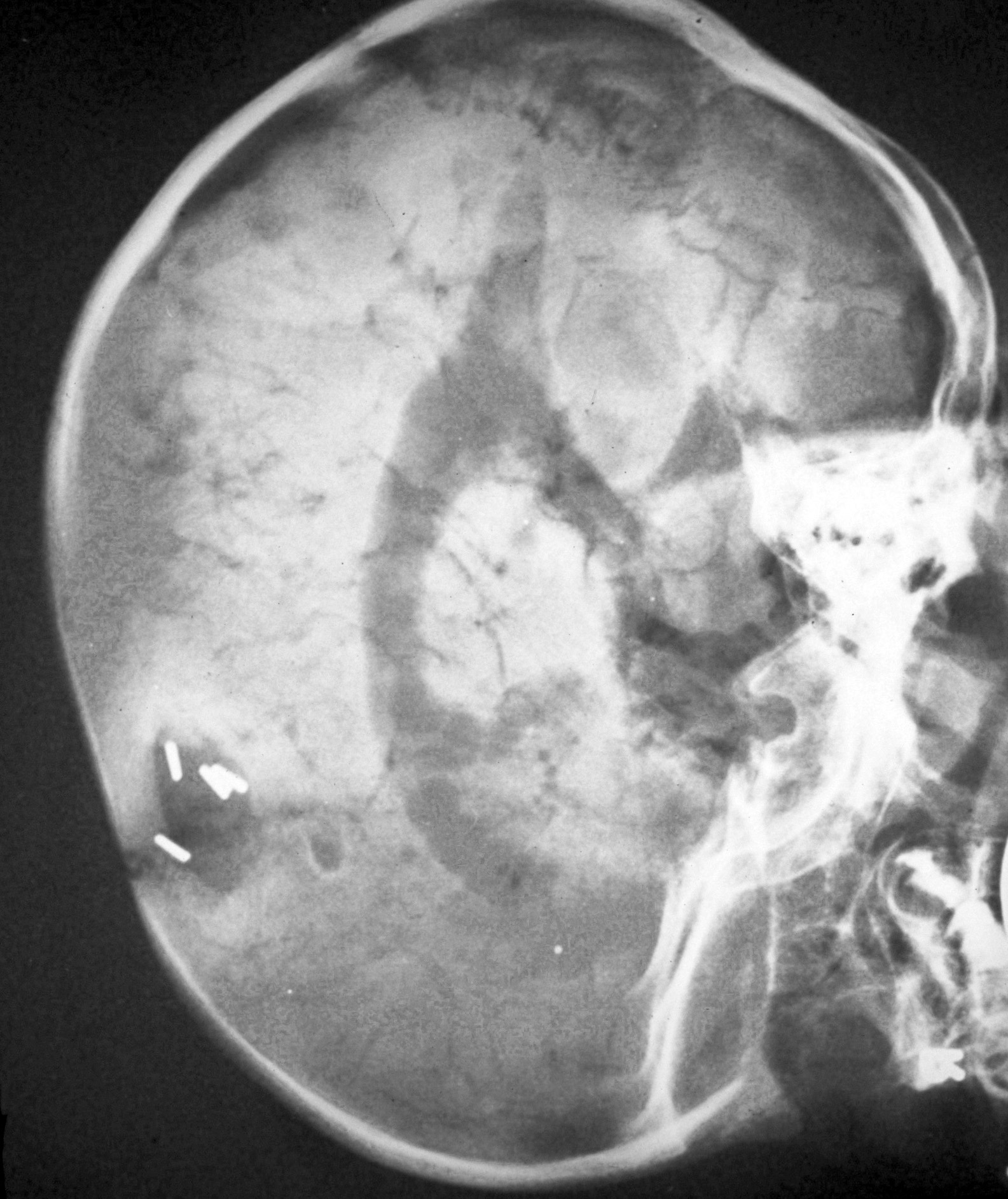
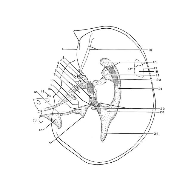
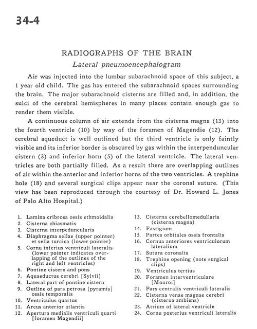
Air was injected into the lumbar subarachnoid space of this subject, a 1 year old child. The gas has entered the subarachnoid spaces surrounding the brain. The major subarachnoid cisterns are filled and, in addition, the sulci of the cerebral hemispheres in many places contain enough gas to render them visible.
- Cribriform plate of ethmoid bone
- Chiasmatic cistern
- Interpeduncular cistern
- Diaphragma sellae (upper pointer) & sella turcica (lower pointer)
- Inferior horn of lateral ventricle (lower pointer indicates overlapping of the outlines of the right and left ventricles)
- Pontine cistern and pons
- Cerebral aqueduct
- Lateral part of pontine cistern
- Outline of petrosal part of temporal bone
- Fourth ventricle
- Anterior arch of atlas
- Medial aperture fourth ventricle (foramen Magendie)
- Cerebellomedullary cistern (cisterna magna)
- Fastigium
- Orbital part of frontal bone
- Anterior horn of lateral ventricle
- Coronal Suture
- Trephine opening (note surgical clips)
- Third ventricle
- Interventricular foramen (of Monro)
- Central part lateral ventricle
- Cisterna venae magnae cerebri (cisterna ambiens)
- Atrium of lateral ventricle
- Posterior horn lateral ventricle