X CLOSE

Exploration of the spinal cord and meninges in situ

Epidural space in thoracic region

Stanford holds the copyright to the David L. Bassett anatomical images and has assigned Creative Commons license Attribution-Share Alike 4.0 International to all of the images.
For additional information regarding use and permissions, please contact Dr. Drew Bourn at dbourn@stanford.edu.

Image #33-2
Bassett ImageDiagram Image
Legend Image
Exploration of the spinal cord and meninges in situ
Epidural space in thoracic region
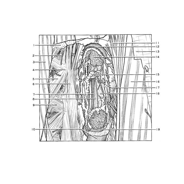
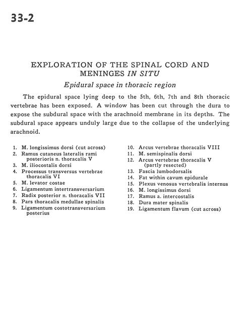
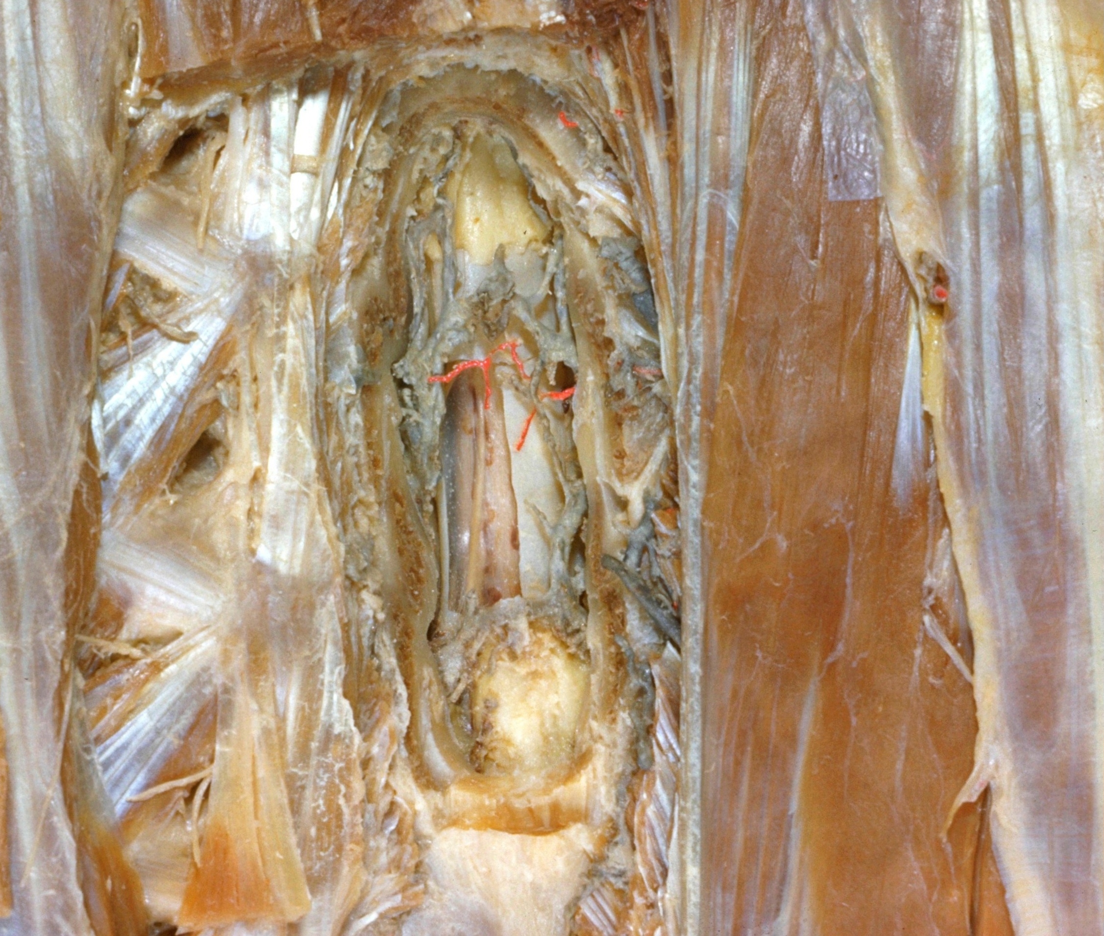
The epidural space lying deep to the 5th, 6th, 7th and 8th thoracic vertebrae has been exposed. A window has been cut through the dura to expose the subdural space with the arachnoid membrane in its depths. The subdural space appears unduly large due to the collapse of the underlying arachnoid.
1 . Longissimus dorsi muscle (cut across)
2 . Lateral cutaneous branch of posterior brach of thoracic nerve V
3 . Dorsal iliocostal muscle
4 . Transverse process thoracic vertebrae VI
5 . Levator costarum muscle
6 . Intertransverse ligament
7 . Dorsal root thoracic nerve VII
8 . Thoracic part spinal cord
9 . Posterior costotransverse ligament
10 . Arch of thoracic vertebra VIII
11 . Dorsal semispinalis muscle
12 . Arch of thoracic vertebra V (partly resected)
13 . Lumbodorsal fascia
14 . Fat within epidural space
15 . Internal vertebral venous plexus
16 . Longissimus dorsi muscle
17 . Branch of intercostal artery
18 . Dura mater
19 . Ligamentum flavum (cut across)