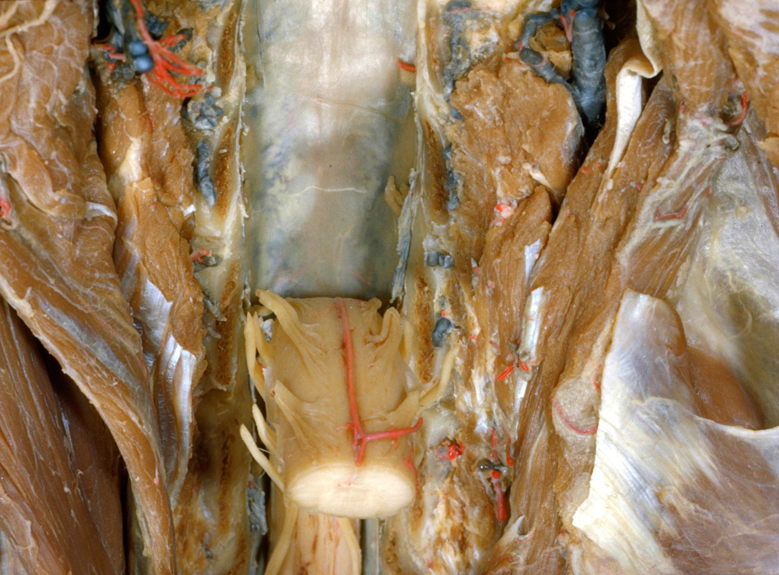
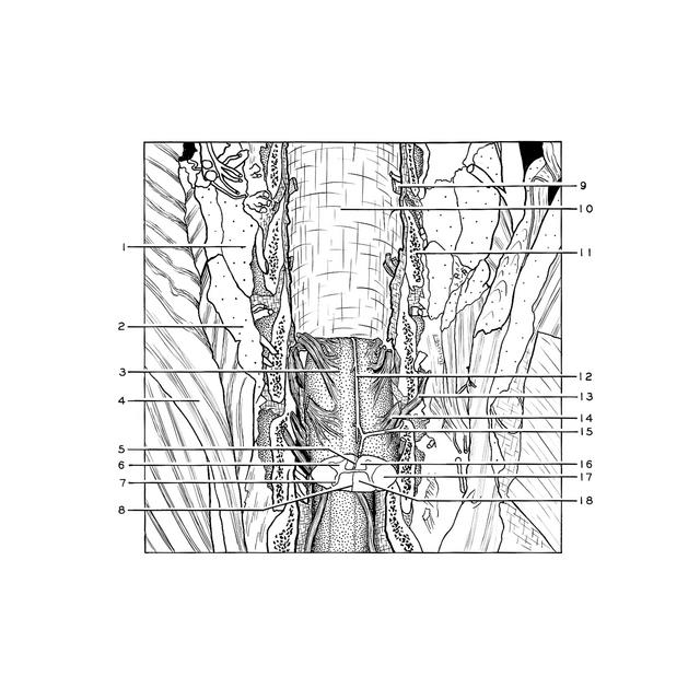
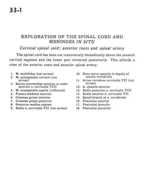
Exploration of the spinal cord and meninges in situ
Cervical spinal cord; anterior roots and spinal artery
Stanford holds the copyright to the David L. Bassett anatomical images and has assigned
Creative Commons license Attribution-Share
Alike 4.0 International to all of the images.
For additional information regarding use and permissions,
please contact Dr. Drew Bourn at dbourn@stanford.edu.
Image #33-1
Exploration of the spinal cord and meninges in situ
Cervical spinal cord; anterior roots and spinal artery
The spinal cord has been cut transversely immediately above the seventh cervical segment and the lower part retracted posteriorly. This affords a view of the anterior roots and anterior spinal artery.
- Multifidus muscle (cut across)
- Semispinalis cervicis muscle (cut across)
- Anterior intermediate sulcus and ventral root cervical nerve VIII
- Semispinalis capitis muscle (reflected)
- Ventral median fissure
- Anterior column (gray matter)
- Posterior column (gray matter)
- Posterior median septum
- Root cervical nerve VII (cut across)
- Dura mater in depths of vertebral canal
- Arch of cervical vertebra VII (cut across)
- Anterior spinal artery
- Dorsal root cervical nerve VIII
- Ventral root cervical nerve VII
- Spinal branch of vertebral artery
- Anterior funiculus
- Lateral funiculus
- Posterior funiculus