X CLOSE

Exploration of the meninges and brain in situ

Rhomboid fossa

Stanford holds the copyright to the David L. Bassett anatomical images and has assigned Creative Commons license Attribution-Share Alike 4.0 International to all of the images.
For additional information regarding use and permissions, please contact Dr. Drew Bourn at dbourn@stanford.edu.

Image #3-7
Bassett ImageDiagram Image
Legend Image
Exploration of the meninges and brain in situ
Rhomboid fossa
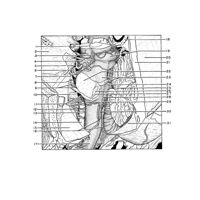
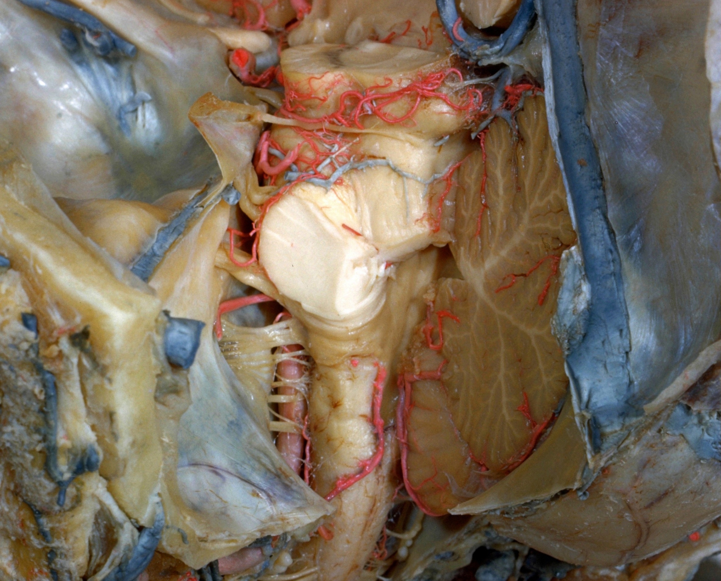
The choroid plexus and tela chorioidea have been removed to expose the entire left half of the fourth ventricle. The inferior margin of the lateral recess of the ventricle is marked by the cut edge of the taenia ventriculi quarti (29) which can be traced laterally across several rootlets of the ninth and tenth nerves.
1 . Cerebral peduncle (cut across)
2 . Middle cranial fossa
3 . Branch of posterior cerebral artery
4 . Trochlear nerve (IV)
5 . Superior cerebellar artery (cut across)
6 . Major portion of trigeminal nerve (V)
7 . Minor portion of trigeminal nerve (V)
8 . Internal auditory artery
9 . Facial nerve (VII) and vestibulocochlear (VIII)
10 . Anterior inferior cerebellar artery (cut across)
11 . Roots glossopharyngeal nerve (IX), vagus (X), and accessory (XI)
12 . Spinal root of accessory nerve
13 . Hypoglossal nerve root (XII) (other rootlets of this nerve also visible)
14 . Vertebral artery left
15 . Posterior cranial fossa
16 . Posterior inferior cerebellar arteries
17 . Spinal medulla
18 . Great cerebral vein (of Galen)
19 . Inferior colliculus
20 . Falx cerebri
21 . Straight sinus (opened)
22 . Brachium pontis (middle cerebellar peduncle) (cut across)
23 . Superior cerebellar peduncle (cut across)
24 . Superior part of rhomboid fossa
25 . Arbor vitae
26 . Inferior cerebellar peduncle(cut across)
27 . Nucleus dorsalis cochlear nerve
28 . Intermediate part of rhomboid fossa
29 . Lateral recess of rhomboid fossa and taenia fourth ventricle
30 . Inferior part of rhomboid fossa
31 . Falx cerebelli