Exploration of the meninges and brain in situ
Relation of trochlear nerve to superior cerebellar artery and tentorium
Stanford holds the copyright to the David L. Bassett anatomical images and has assigned
Creative Commons license Attribution-Share
Alike 4.0 International to all of the images.
For additional information regarding use and permissions,
please contact Dr. Drew Bourn at dbourn@stanford.edu.
Image #3-3
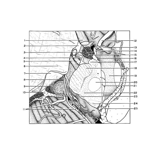
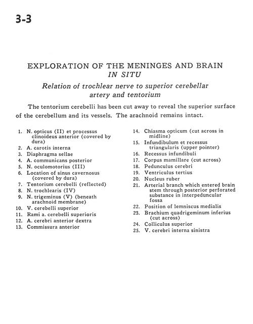
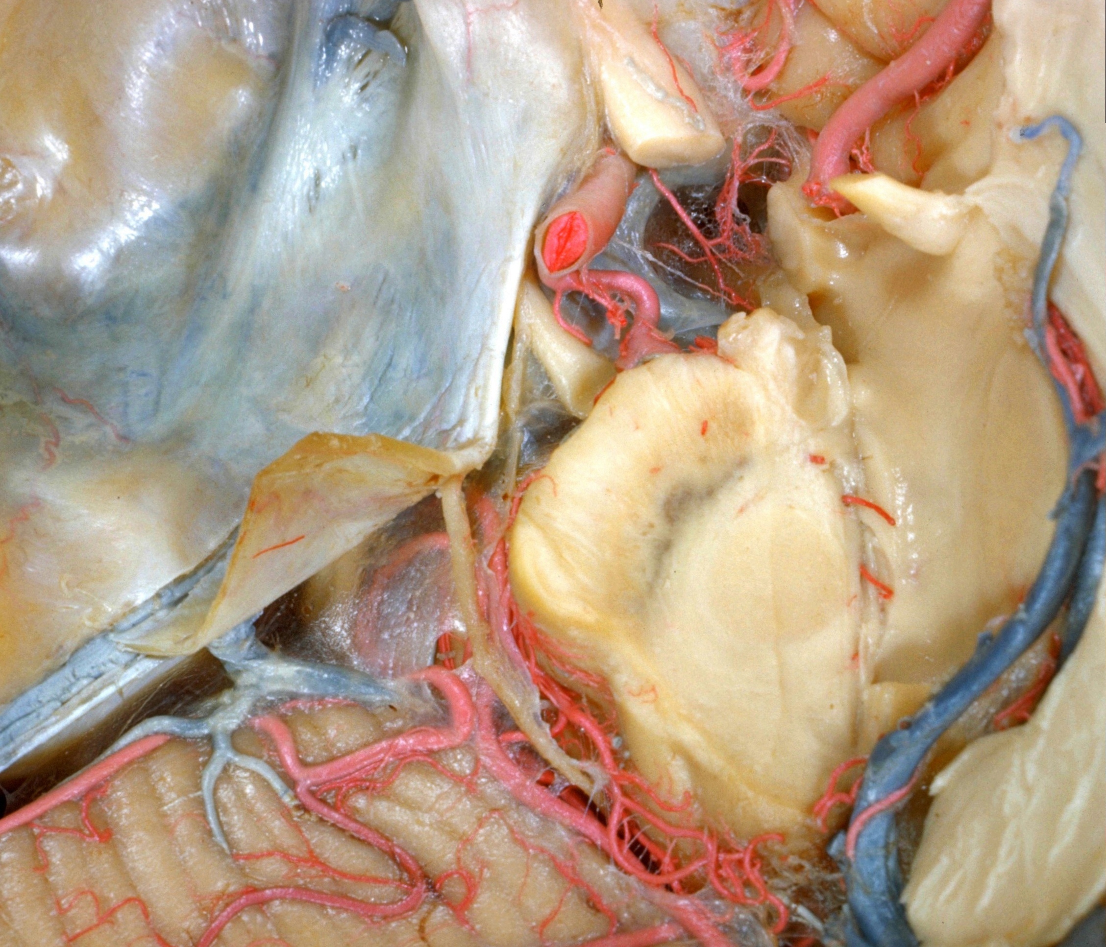
Exploration of the meninges and brain in situ
Relation of trochlear nerve to superior cerebellar artery and tentorium
The tentorium cerebelli has been cut away to reveal the superior surface of the cerebellum and its vessels. The arachnoid remains intact.
- Optic nerve (II) and anterior clinoid process (covered by dura)
- Internal carotid artery
- Diaphragma sellae
- Posterior communicating artery
- Oculomotor nerve (III)
- Location of cavernous sinus (covered by dura)
- Tentorium cerebelli (reflected)
- Trochlear nerve (IV)
- Trigeminal nerve (V) (beneath arachnoid membrane)
- Superior cerebellar vein
- Branch superior cerebellar artery
- Anterior cerebral artery right
- Anterior commissure
- Optic chiasm (cut across in midline)
- Infundibulum and triangular recess (upper pointer)
- Infundibular recess
- Mamillary body (cut across)
- Cerebral peduncle
- Third ventricle
- Red nucleus
- Arterial branch which entered brain stem through posterior perforated substance in interpeduncular fossa
- Position of medial lemniscus
- Brachium inferior quadrigeminal (cut across)
- Superior colliculus
- Internal cerebral vein left