X CLOSE

Exploration of the left half of the cerebellum and brain stem from its inferior and medial aspect

Continuity of medullary center of tonsil with uvula; peduncle of flocculus

Stanford holds the copyright to the David L. Bassett anatomical images and has assigned Creative Commons license Attribution-Share Alike 4.0 International to all of the images.
For additional information regarding use and permissions, please contact Dr. Drew Bourn at dbourn@stanford.edu.

Image #26-3
Bassett ImageDiagram Image
Legend Image
Exploration of the left half of the cerebellum and brain stem from its inferior and medial aspect
Continuity of medullary center of tonsil with uvula; peduncle of flocculus
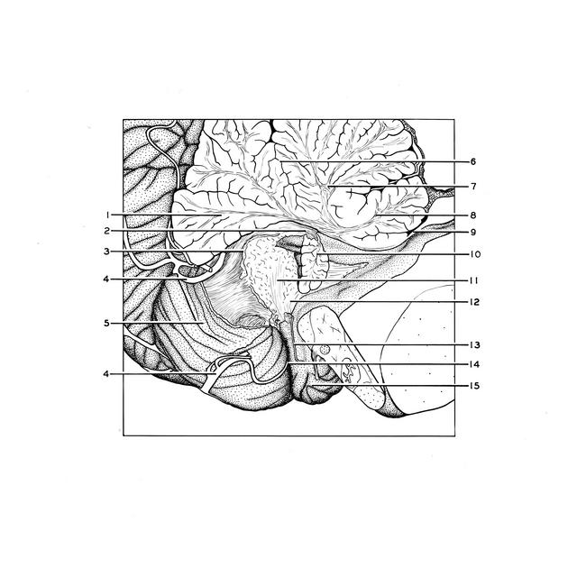
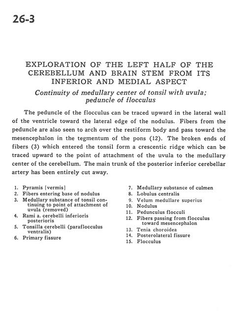
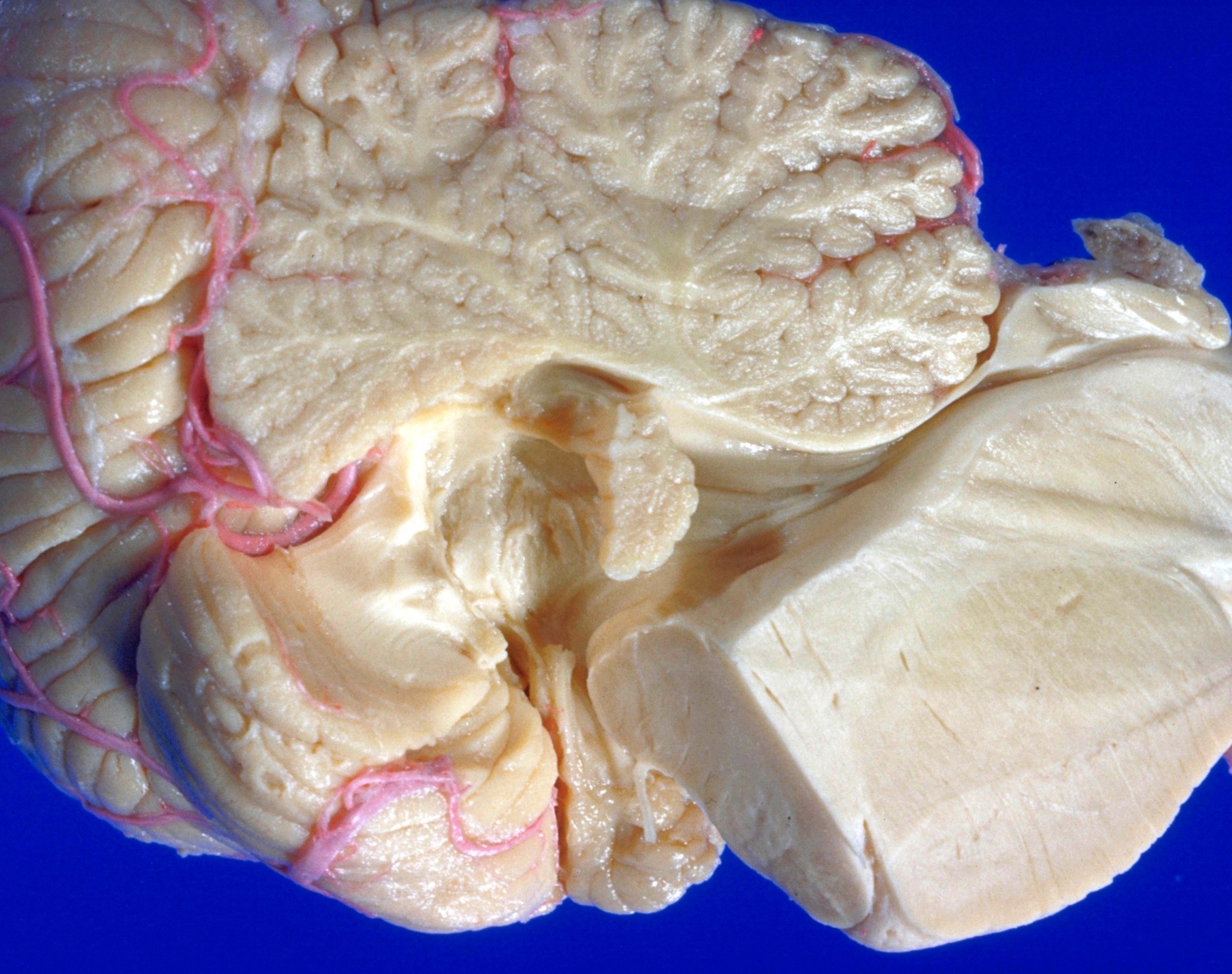
The peduncle of the flocculus can be traced upward in the lateral wall of the ventricle toward the lateral edge of the nodulus. Fibers from the peduncle are also seen to arch over the restiform body and pass toward the mesencephalon in the tegmentum of the pons (12). The broken ends of fibers (3) which entered the tonsil form a crescentic ridge which can be traced upward to the point of attachment of the uvula to the medullary center of the cerebellum. The main trunk of the posterior inferior cerebellar artery has been entirely cut away.
1 . Pyramid (vermis)
2 . Fibers entering base of nodulus
3 . Medullary substance of tonsil continuing to point of attachment of uvula (removed)
4 . Branch of posterior inferior cerebellar artery (PICA)
5 . Cerebellar tonsil (ventral paraflocculus)
6 . Primary fissure
7 . Medullary substance of culmen
8 . Central lobule
9 . Anterior medullary velum
10 . Nodulus
11 . Floccular peduncle
12 . Fibers passing from flocculus toward mesencephalon
13 . Taenia chorioidea
14 . Posterolateral fissure
15 . Flocculus