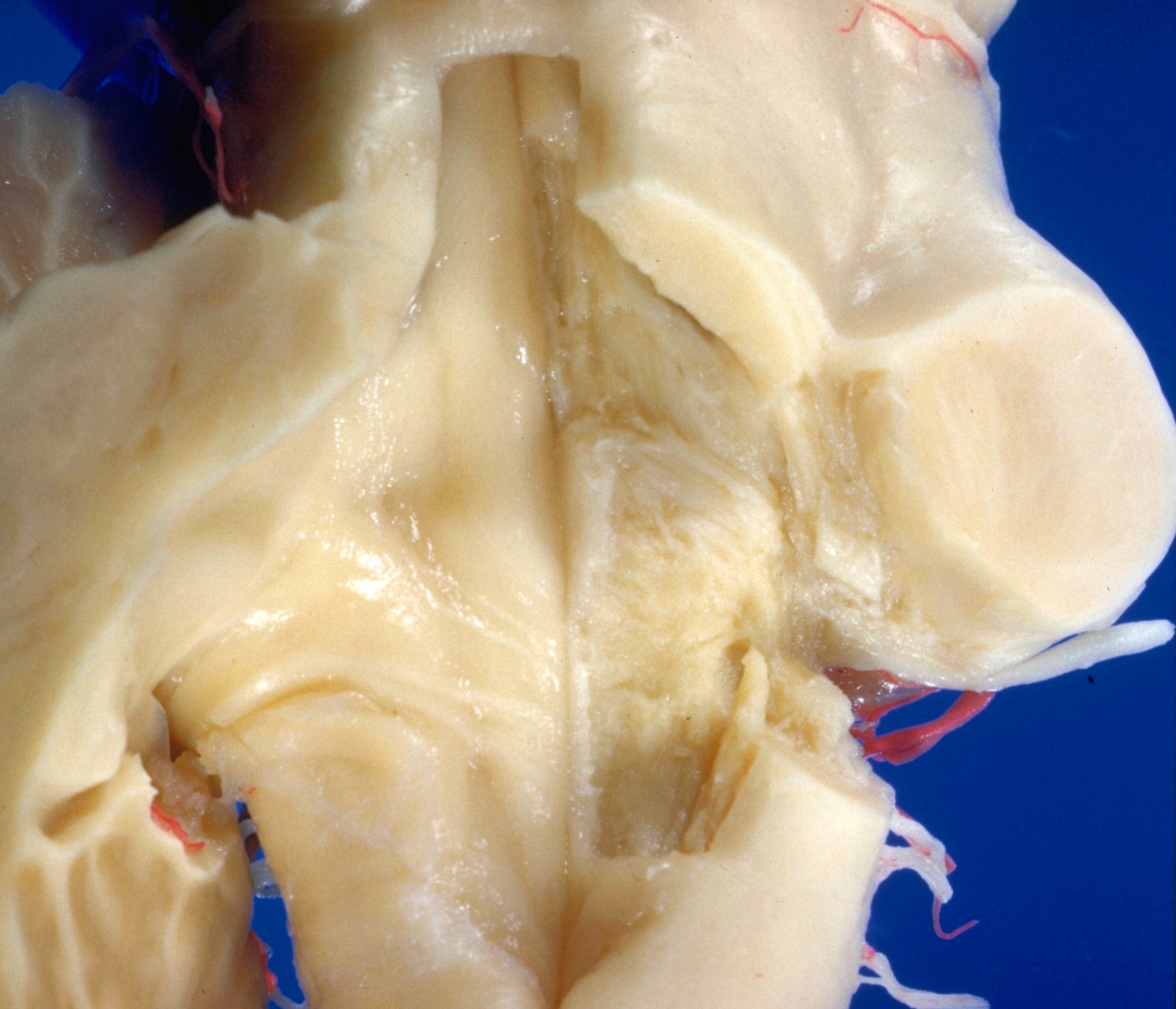
Exploration of the cerebellum from above and behind
Internal genu of facial nerve, nucleus of abducens nerve and tractus solitarius
Stanford holds the copyright to the David L. Bassett anatomical images and has assigned
Creative Commons license Attribution-Share
Alike 4.0 International to all of the images.
For additional information regarding use and permissions,
please contact Dr. Drew Bourn at dbourn@stanford.edu.
Image #25-1
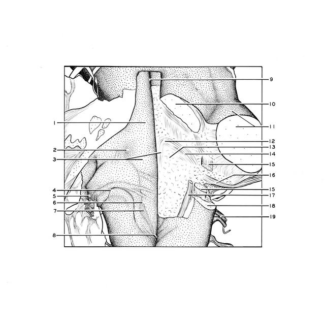
Exploration of the cerebellum from above and behind
Internal genu of facial nerve, nucleus of abducens nerve and tractus solitarius
The floor of the rhomboid fossa has been dissected on the right side to expose the internal genu of the facial nerve (12) which passes around the nucleus of the abducens nerve (13). The spinal tract of the trigeminal nerve (15) has been divided to expose the root of the facial nerve (14) as it continues toward the point of exit of the facial nerve (16) behind the brachium pontis.
- Median eminence rhomboid fossa
- Superior fovea rhomboid fossa
- Origin of roots of facial nerve and facial colliculus
- Taenia fourth ventricle
- Lateral recess of rhomboid fossa
- Inferior fovea rhomboid fossa
- Wing of gray matter
- Calamus scriptorius
- Median sulcus of rhomboid fossa
- Brachium conjunctivum (superior cerebellar peduncle) (cut across)
- Brachium pontis (middle cerebellar peduncle) (cut across)
- Genu internal roots of facial nerve
- Nucleus abducens nerve
- Second part roots of facial nerve
- Spinal trigeminal tract (divided)
- Facial nerve
- Tractus solitarius
- Restiform body (inferior cerebellar peduncle) (cut across)
- Vagus nerve (X)