Dissection of anterior aspect of vertebral column
Thoracic region.
Stanford holds the copyright to the David L. Bassett anatomical images and has assigned
Creative Commons license Attribution-Share
Alike 4.0 International to all of the images.
For additional information regarding use and permissions,
please contact Dr. Drew Bourn at dbourn@stanford.edu.
Image #220-5
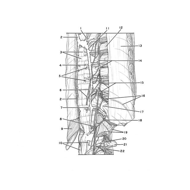
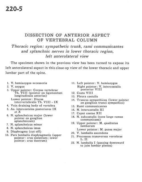
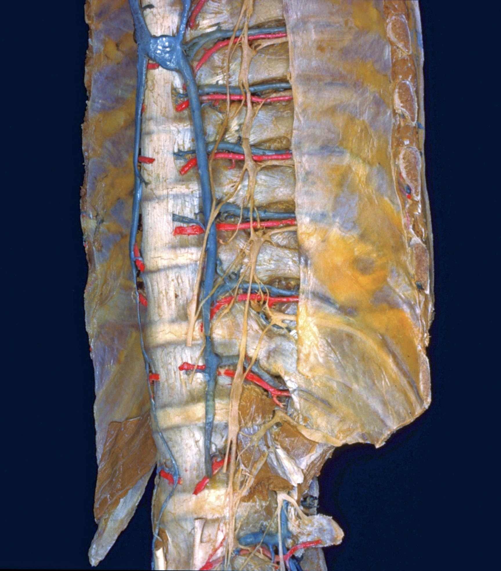
Dissection of anterior aspect of vertebral column
Thoracic region.
The specimen shown in the previous view has been turned to expose its left anterolateral aspect in this close-up view of the lower thoracic and upper lumbar part of the spine.
- Accessory hemiazygos vein
- Azygos vein
- Upper pointer: Body of vertebra Th. VIII (pointer on anterior longitudinal ligament) Lower pointer: Intervertebral disc Th. VII-IX
- Vein draining body of vertebra
- Posterior intercostal arteries IX and X
- Greater splanchnic nerve (lower pointer on splanchnic ganglion)
- Lesser splanchnic nerve
- Least splanchnic it
- Diaphragm (cut off)
- Lumbar part of diaphragm (upper pointer: left crus; lower pointer: right crus)
- Left pointer: Hemiazygos vein Right pointer: Posterior intercostal vein VIII
- Rib VIII
- Costal pleura
- Sympathetic trunk (lower pointer on ganglion of sympathetic trunk)
- Rami communicantes
- Intercostal nerve XI
- Head of rib XII
- Subcostal nerve (note large ramus communicans)
- Upper pointer: Quadratus lumborum muscle Lower pointer: Psoas major muscle
- Ascending lumbar vein
- Transverse process of vertebra L. II
- Lumbar nerve I (passing downward to join lumbar plexus)