X CLOSE

Thoracic meninges, spinal cord and nerve roots dissected in relation to vertebral column

Intervertebral course and meningeal branches of left fifth thoracic nerve

Stanford holds the copyright to the David L. Bassett anatomical images and has assigned Creative Commons license Attribution-Share Alike 4.0 International to all of the images.
For additional information regarding use and permissions, please contact Dr. Drew Bourn at dbourn@stanford.edu.

Image #219-1
Bassett ImageDiagram Image
Legend Image
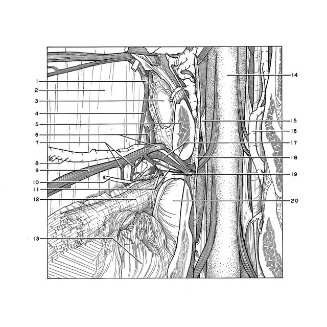
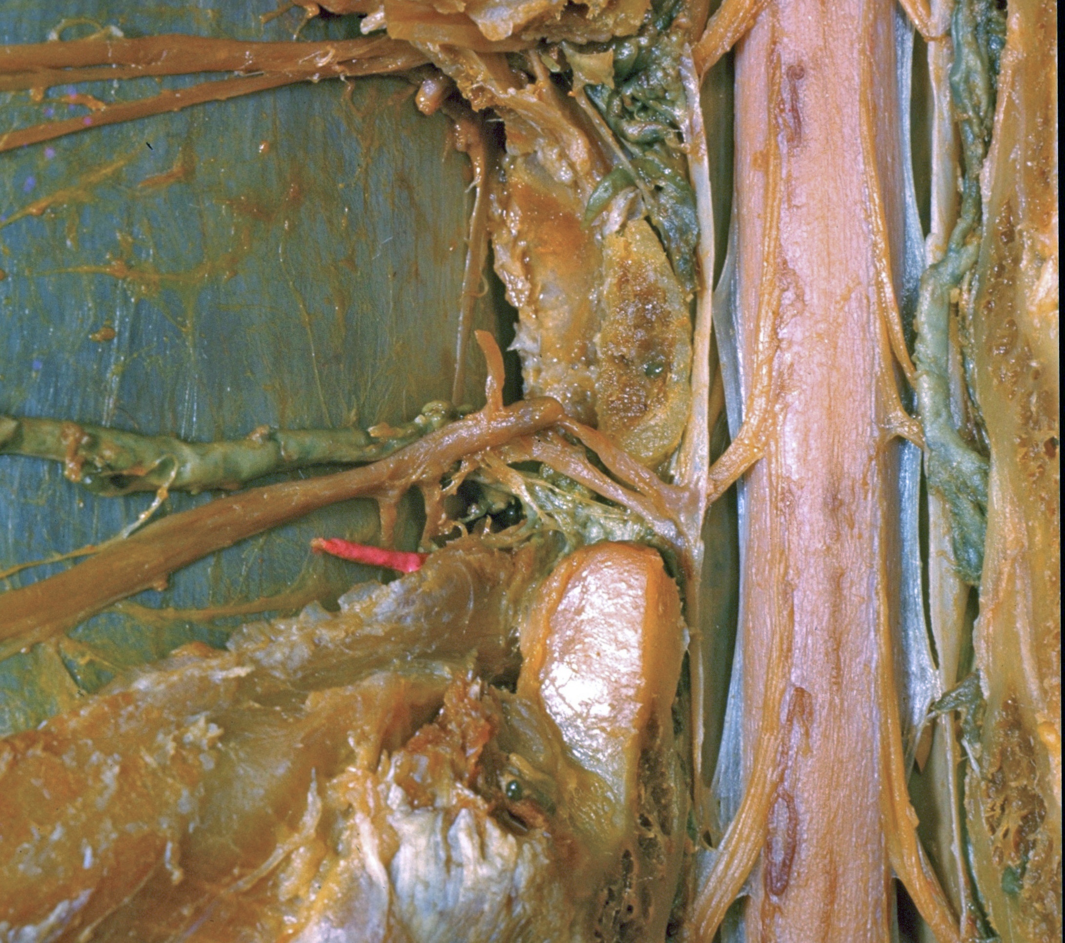
Thoracic meninges, spinal cord and nerve roots dissected in relation to vertebral column
Intervertebral course and meningeal branches of left fifth thoracic nerve
The fifth rib has been removed from the left side. The vertebral canal has been opened by laminectomy and the pedicle of the fifth thoracic vertebra has been cut off on the left to expose the component parts of the corresponding spinal nerve. A plexus of vertebral branches (11) of this nerve, including meningeal filaments, can be seen to extend medially through the intervertebral foramen anterior to the nerve roots which have been elevated somewhat out of their usual position.
1 . Thoracic sympathetic ganglion
2 . Costal pleura
3 . Joint head of rib V (rib removed)
4 . Sympathetic trunk
5 . Pedicle (arch of vertebra) Th. V (cut off)
6 . Upper pointer: Dorsal branch thoracic nerve V Lower pointer: Spinal ganglion thoracic nerve V
7 . Rami communicantes
8 . Posterior intercostal veins
9 . Intercostal nerve V
10 . Posterior intercostal artery V
11 . Meningeal branch thoracic nerve V
12 . Neck of rib (superficial crest rib visible above pointer on upper margin of rib)
13 . Transverse process of vertebra Th. VI
14 . Spinal cord
15 . Denticulate ligament
16 . Posterior internal vertebral venous plexus
17 . Left pointer: Ventral root thoracic nerve V Right pointer: Dorsal roots thoracic nerve V
18 . Dura mater
19 . Intervertebral foramen
20 . Superior articular surface