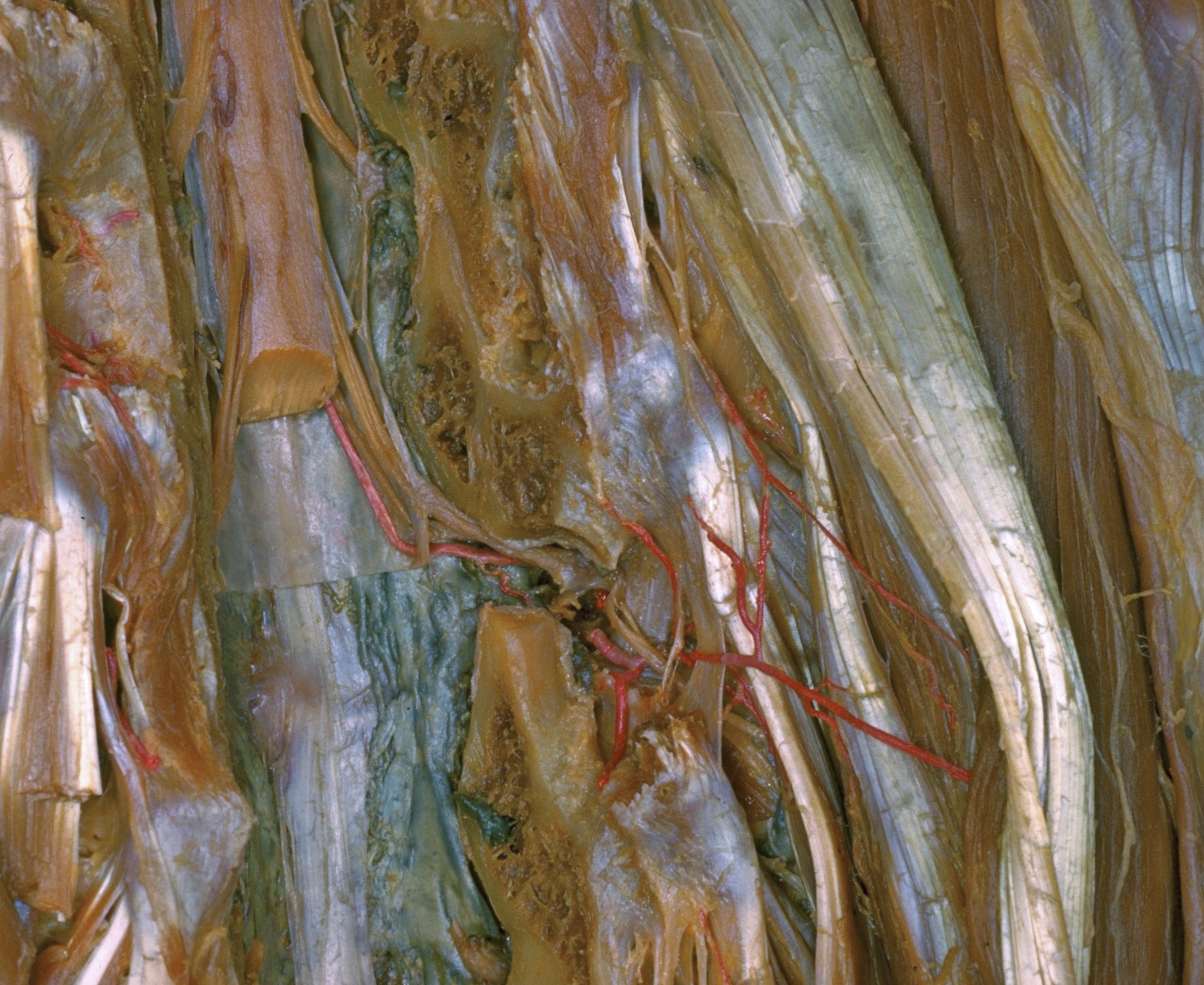
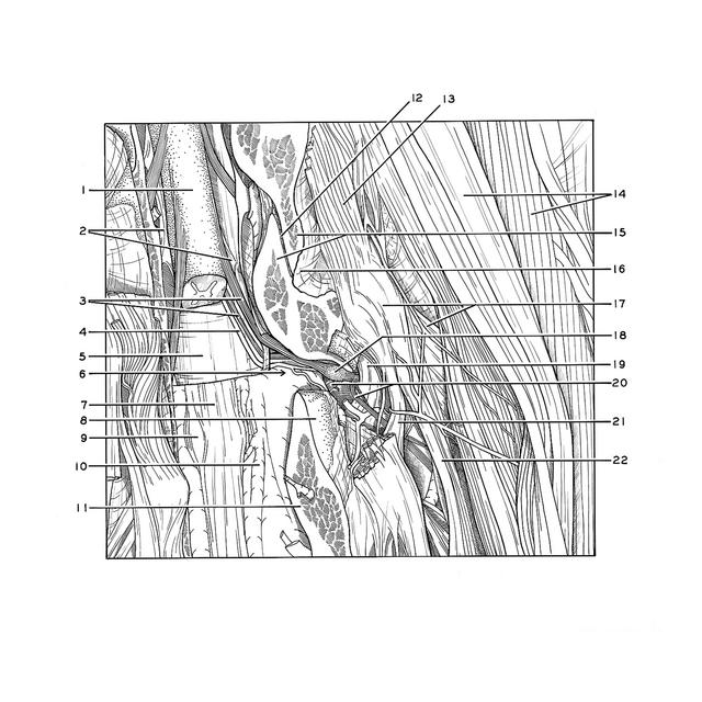
Thoracic meninges, spinal cord and nerve roots dissected in relation to vertebral column
Right tenth thoracic nerve within vertebral canal and intervertebral foramen
Stanford holds the copyright to the David L. Bassett anatomical images and has assigned
Creative Commons license Attribution-Share
Alike 4.0 International to all of the images.
For additional information regarding use and permissions,
please contact Dr. Drew Bourn at dbourn@stanford.edu.
Image #218-7
Thoracic meninges, spinal cord and nerve roots dissected in relation to vertebral column
Right tenth thoracic nerve within vertebral canal and intervertebral foramen
The vertebral canal has been opened by laminectomy and the tenth intervertebral foramen (6) has been opened on the right side by excision of the inferior articular process of the tenth thoracic vertebra. The dorsal and ventral roots of the corresponding nerve have been exposed. The roots penetrate the dura separately in this specimen.
- Spinal cord
- Denticulate ligaments
- Upper pointer: Dorsal roots thoracic nerve X Lower pointer: Ventral root thoracic nerve X
- Spinal branch posterior intercostal artery
- Arachnoid and dura mater
- Intervertebral foramen (opened venous plexus partially resected)
- Posterior longitudinal ligament
- Articular surface
- Protrusion of intervertebral disc
- Anterior internal vertebral venous plexus
- Pedicle (arch of vertebra) Th. Xl
- Articular cavity of intervertebral joint
- Intertransverse thoracis muscle
- Left pointer: Longissimus thoracis muscle Right pointer: Iliocostalis muscle
- Upper pointer: Inferior articular process vertebra Th. IX Lower pointer: Superior articular process vertebra Th. IX
- Joint capsule
- Left pointer: Tranverse process vertebra Th. X Right pointer: Levator costarum brevis muscle
- Spinal ganglion thoracic nerve X
- Superior costotransverse ligament
- Upper pointer: Ramus communicans Lower pointer: Dorsal branch thoracic nerve X
- Intertransverse ligament
- Longissimus thoracis muscle (tendon of insertion)