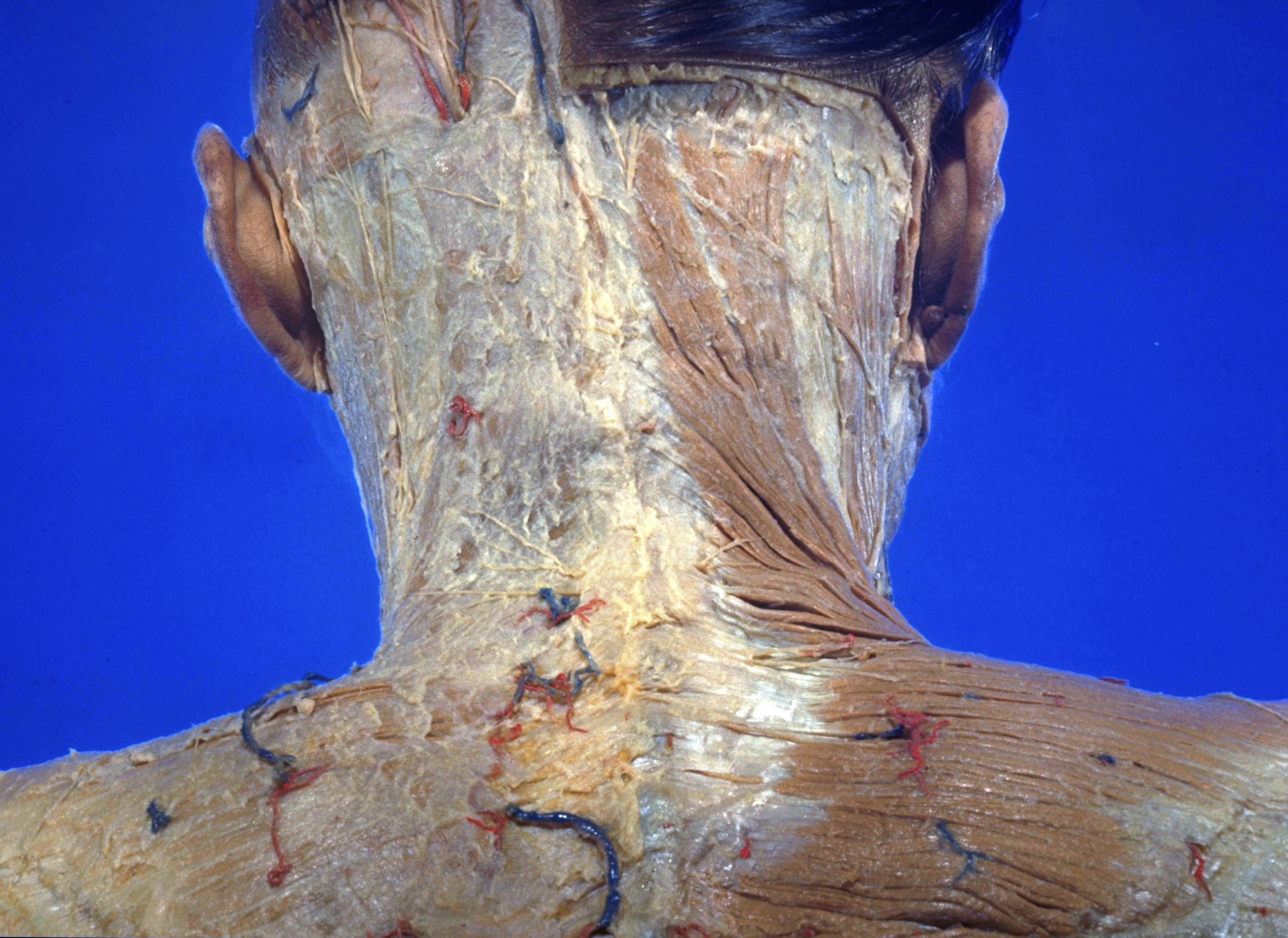
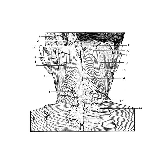
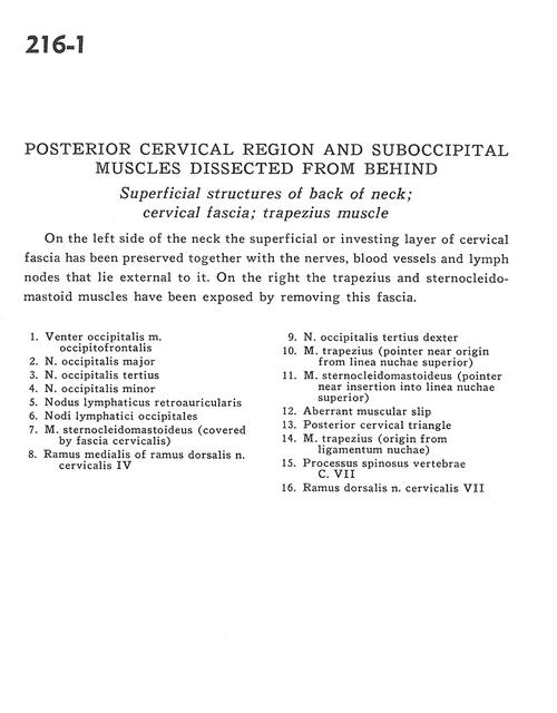
Posterior cervical region and suboccipital muscles dissected from behind
Superficial structures of back of neck; cervical fascia; trapezius muscle
Stanford holds the copyright to the David L. Bassett anatomical images and has assigned
Creative Commons license Attribution-Share
Alike 4.0 International to all of the images.
For additional information regarding use and permissions,
please contact Dr. Drew Bourn at dbourn@stanford.edu.
Image #216-1
Posterior cervical region and suboccipital muscles dissected from behind
Superficial structures of back of neck; cervical fascia; trapezius muscle
On the side of the neck the superficial or investing layer of cervical fascia has been preserved together with the nerves, blood vessels and lymph nodes that lie external to it. On the right the trapezius and sternocleidomastoid muscles have been exposed by removing this fascia
- Occipital belly of occipitofrontal muscle
- Greater occipital nerve
- Third occipital nerve
- Lesser occipital nerve
- Retroauricular lymph node
- Occipital lymph node
- Sternocleidomastoid muscle (covered by cervical fascia)
- Medial branch of dorsal branch cervical nerve IV
- Third occipital nerve right
- Trapezius muscle (pointer near origin from superior nuchal line)
- Sternocleidomastoid muscle (pointer near insertion into superior nuchal line)
- Aberrant muscular slip
- Posterior cervical triangle
- Trapezius muscle (origin from nuchal ligament)
- Spinous process vertebra C. VII
- Dorsal branch cervical nerve VII