Ligaments and joints of lumbosacral spine and pelvic girdle
Ligaments of lumbosacral region, posterior view
Stanford holds the copyright to the David L. Bassett anatomical images and has assigned
Creative Commons license Attribution-Share
Alike 4.0 International to all of the images.
For additional information regarding use and permissions,
please contact Dr. Drew Bourn at dbourn@stanford.edu.
Image #214-7
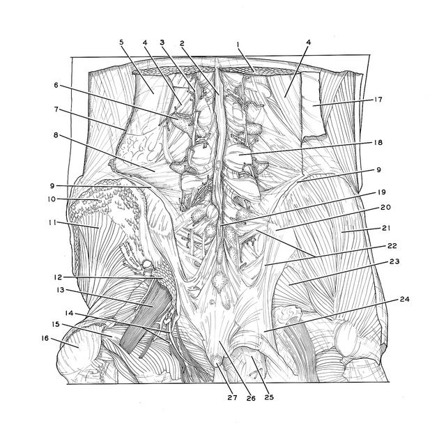
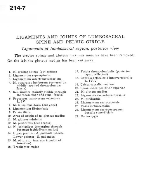
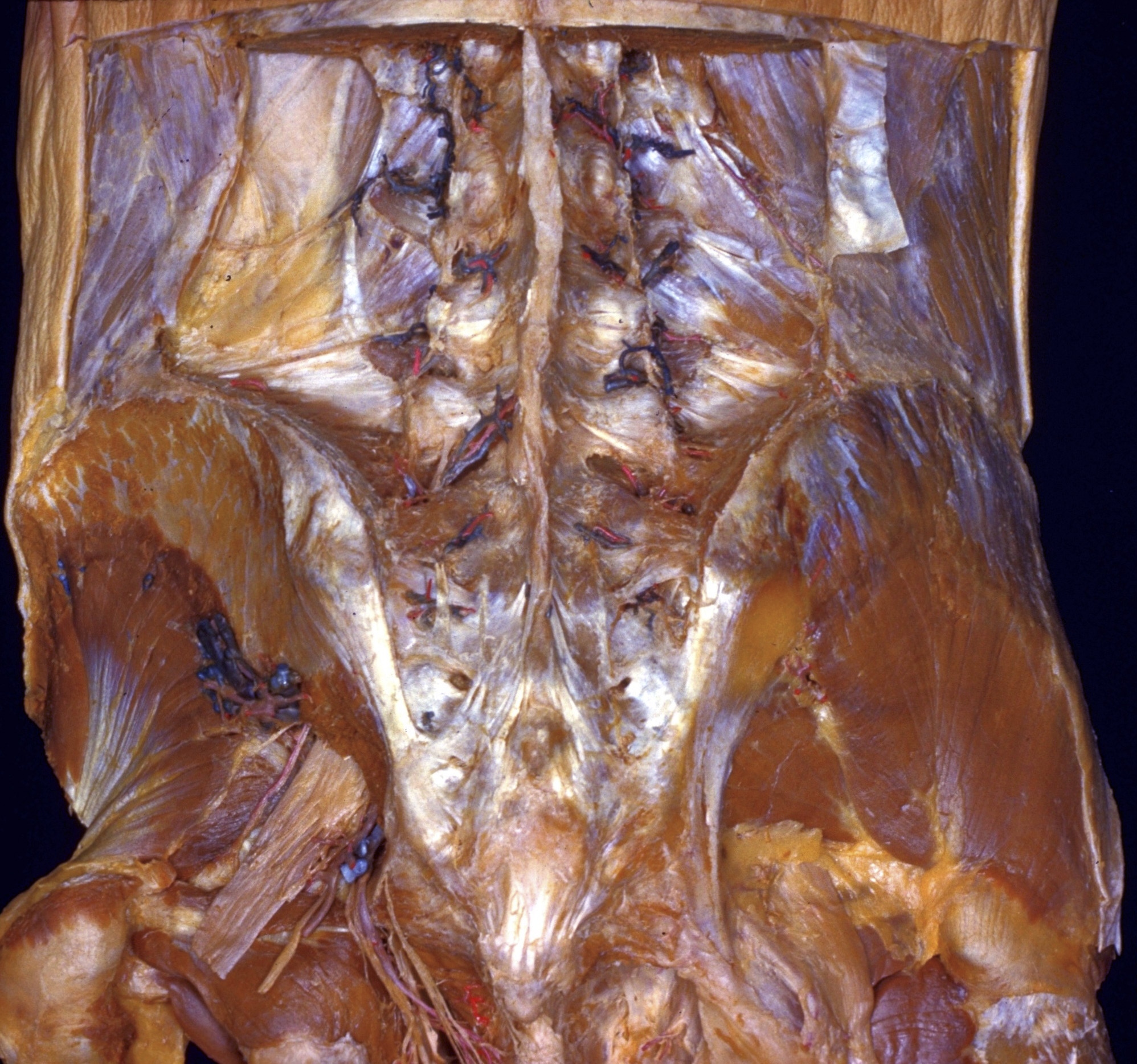
Ligaments and joints of lumbosacral spine and pelvic girdle
Ligaments of lumbosacral region, posterior view
The erector spinae and gluteus maximus muscles have been removed. On the left the gluteus medius has been cut away.
- Erector spinae muscle (cut across)
- Supraspinous ligament
- Intertransverse ligament
- Quadratus lumborum muscle (covered by middle layer of thoracolumbar fascia)
- Left kidney (faintly visible through thoracolumbar and renal fascia)
- Transverse process of vertebra L. IV
- Latissimus dorsi muscle (cut edge)
- Iliolumbar ligament
- Iliac crest
- Area of origin of gluteus medius muscle
- Gluteus minimus muscle
- Piriform muscle (cut across)
- Sciatic nerve (emerging through greater sciatic foramen)
- Upper pointer: Internal pudendal artery Lower pointer: Pudendal nerve
- Obturator internus muscle (tendon of insertion)
- Greater trochanter of femur
- Thoracolumbar fascia (posterior layer, reflected)
- Intervertebral joint capsule L. IV-V
- Median sacral crest
- Posterior superior iliac spine
- Gluteus medius muscle
- Dorsal sacroiliac ligament
- Piriform muscle
- Sacrotuberous ligament
- Ischiorectal fossa
- Dorsal superficial sacrococcygeal ligament
- Coccyx