Dissection of thoracic and lumbosacral regions of back from a posterior approach
Components of erector spinae muscle separated, general view
Stanford holds the copyright to the David L. Bassett anatomical images and has assigned
Creative Commons license Attribution-Share
Alike 4.0 International to all of the images.
For additional information regarding use and permissions,
please contact Dr. Drew Bourn at dbourn@stanford.edu.
Image #213-5
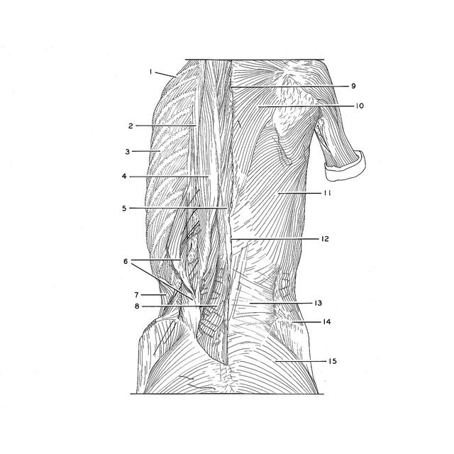
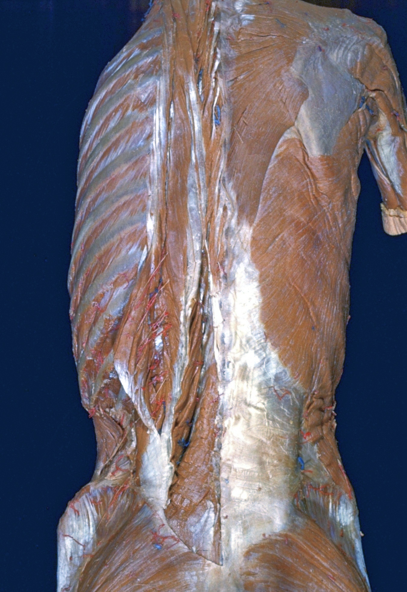
Dissection of thoracic and lumbosacral regions of back from a posterior approach
Components of erector spinae muscle separated, general view
The heavy aponeurosis of origin of the left erector spinae has been partially removed near the midline. This portion of the aponeurosis, form which the longissimus thoracic part arises, also serves to give attachment to portions of the underlying multifidus muscle (8). The iliocostalis (2, 6), longissimus (4) and spinalis (5) muscles have been separated from each other.
- Rib II
- Iliocostalis muscle
- Rib VI
- Longissimus thoracis muscle
- Spinalis thoracis muscle
- Iliocostalis lumborum muscle
- Internal oblique muscle
- Multifidus muscle
- Spinous process vertebra Th. III
- Trapezius muscle
- Latissimus dorsi muscle
- Spinous process vertebra Th. XII
- Thoracolumbar fascia (posterior layer)
- Iliac crest
- Gluteus maximus muscle