X CLOSE

Exploration of those parts of the brain supplied by the posterior cerebral artery

Sublenticular part of internal capsule

Stanford holds the copyright to the David L. Bassett anatomical images and has assigned Creative Commons license Attribution-Share Alike 4.0 International to all of the images.
For additional information regarding use and permissions, please contact Dr. Drew Bourn at dbourn@stanford.edu.

Image #21-1
Bassett ImageDiagram Image
Legend Image
Exploration of those parts of the brain supplied by the posterior cerebral artery
Sublenticular part of internal capsule
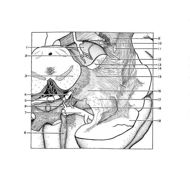
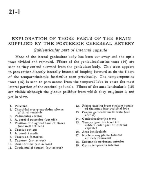
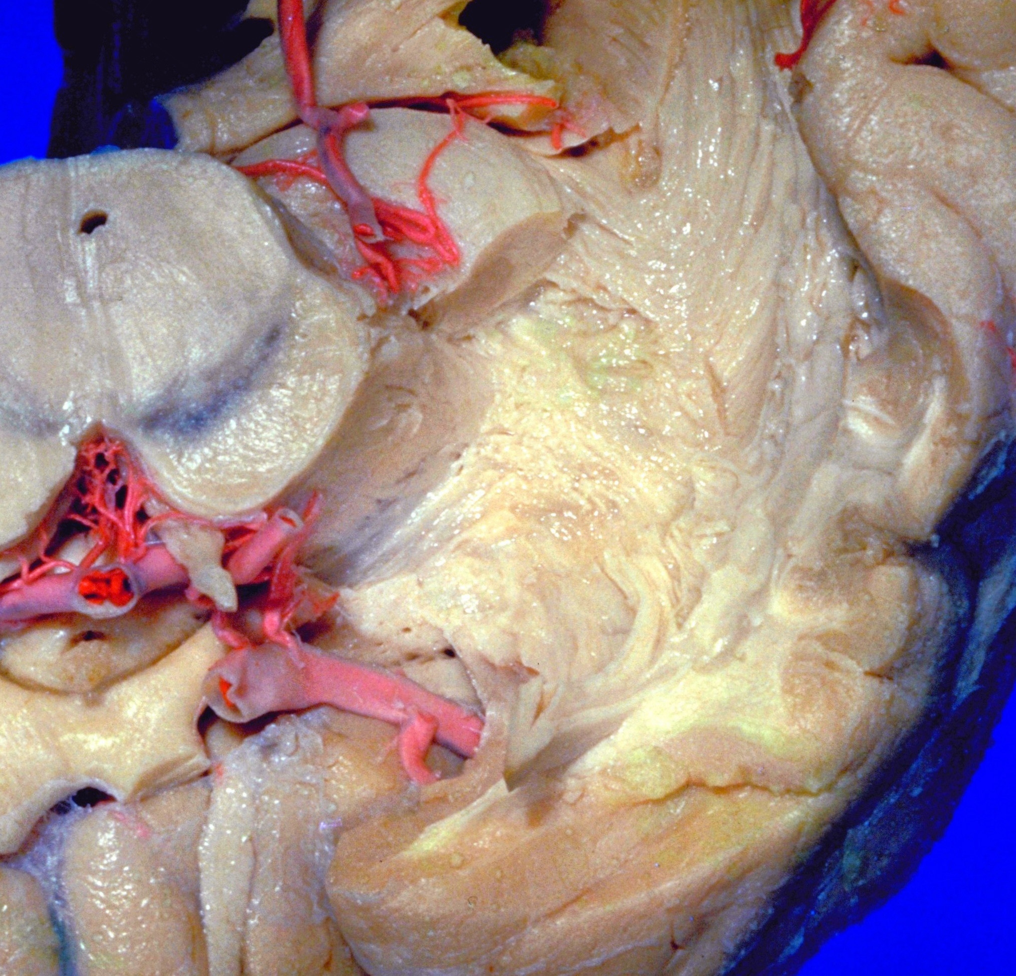
More of the lateral geniculate body has been cut away and the optic tract divided and removed. Fibers of the geniculocalcarine tract (14) are seen as they extend outward from the geniculate body. This tract appears to pass rather directly laterally instead of looping forward as do the fibers of the temporothalamic fasciculus seen previously. The temporopontine tract (15) is seen to pass across from the temporal lobe to enter the most lateral portion of the cerebral peduncle. Fibers of the ansa lenticularis (16) are visible although the globus pallidus from which they originate is not yet in view.
1 . Pulvinar
2 . Choroidal artery supplying plexus of third ventricle
3 . Cerebral peduncle
4 . Posterior cerebral artery (cut off)
5 . Position of diagonal band of Broca (not well defined)
6 . Optic tract
7 . Middle cerebral artery
8 . Olfactory tract
9 . Tapetum (cut across)
10 . Fornix (crus) (cut across)
11 . Caudate nucleus (tail) (cut across)
12 . Fibers passing from stratum zonale of thalamus into occipital lobe
13 . Lateral geniculate body (cut across)
14 . Geniculocalcarine tract
15 . Temporopontine tract (in sublenticular part of internal capsule)
16 . Ansa lenticularis
17 . Amygdaloid nucleus (almost entirely removed)
18 . Anterior perforated substance
19 . Inferior temporal gyrus