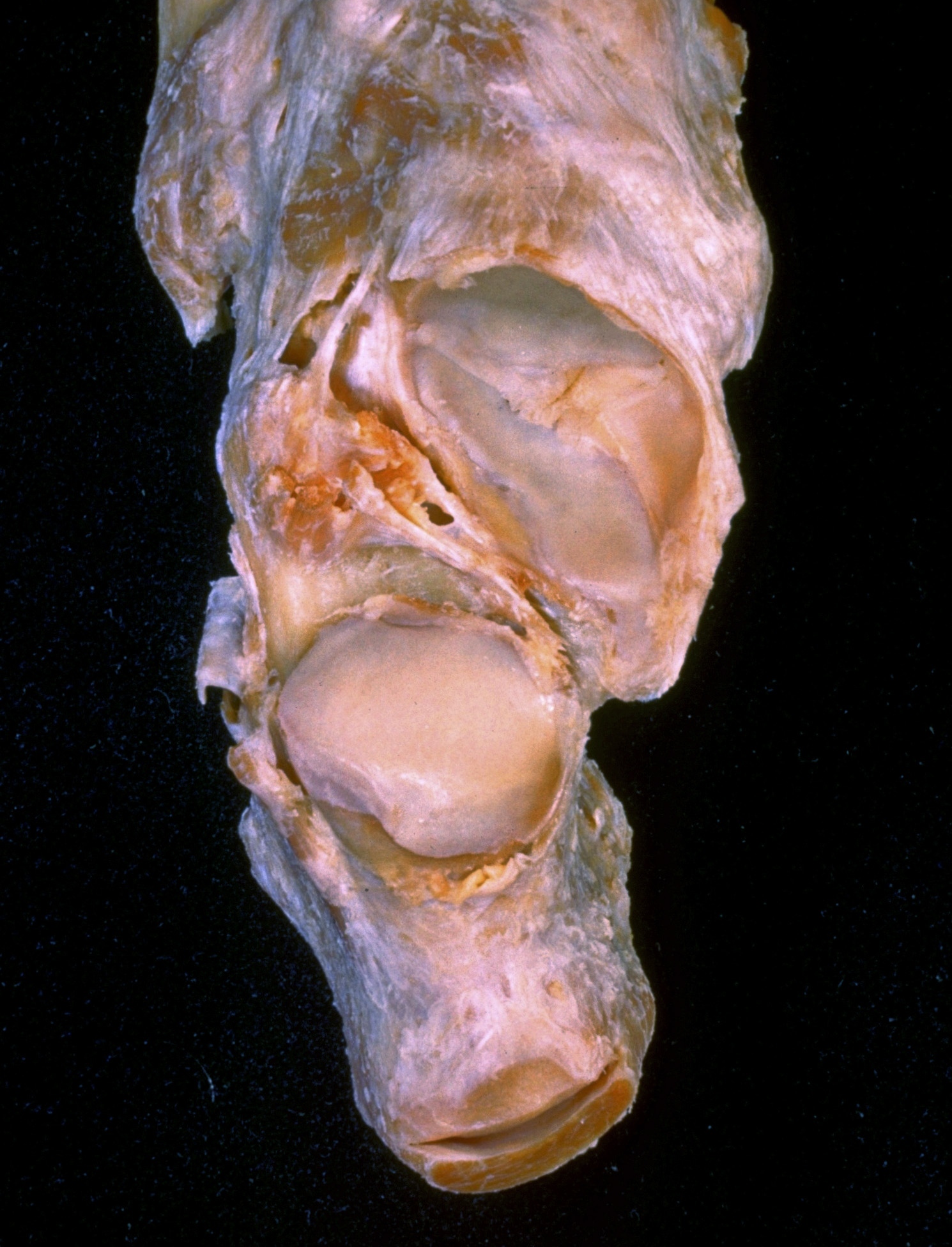
Joints of left ankle and foot
Subtalar and talocalcaneonavicular joints, opened and viewed from above
Stanford holds the copyright to the David L. Bassett anatomical images and has assigned
Creative Commons license Attribution-Share
Alike 4.0 International to all of the images.
For additional information regarding use and permissions,
please contact Dr. Drew Bourn at dbourn@stanford.edu.
Image #204-1
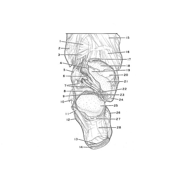
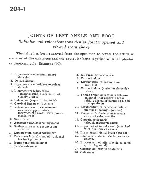
Joints of left ankle and foot
Subtalar and talocalcaneonavicular joints, opened and viewed from above
The talus has been removed from the specimen to reveal the articular surfaces of the calcaneus and the navicular bone together with the plantar calcaneonavicular ligament (20).
- Dorsal cuneonavicular ligament
- Cuboid bone
- Dorsal cuboideonavicular ligament
- Bifurcate ligament (calcaneocuboid ligament not clearly visible)
- Calcaneus (superior tubercle)
- Cervical ligament (cut off)
- Inferior extensor retinaculum (upper pointer: intermediate root; lower pointer: medial root)
- Tarsal sinus
- Anterior talocalcaneal ligament
- Inferior peroneal retinaculum
- Calcaneofibular ligament
- Lateral tubercle process of calcaneus bone (in background)
- Calcaneal subtendinous bursa
- Tendo calcaneus
- Medial cuneiform bone
- Navicular bone
- Talonavicular ligament (cut off)
- Navicular bone (articular surface for talus)
- Anterior articular surface of calcaneus for talus (not separate from middle articular surface (21) in this specimen)
- Plantar calcaneonavicular ligament (spring ligament)
- Medial articular surface of calcaneus for talus (also see 19)
- Talocalcaneonavicular articular capsule
- Ligament of tarsal canal (attached within calcaneal groove)
- Deltoid ligament (cut off)
- Posterior articular surface of calcaneus for talus
- Medial process of tuberosity of calcaneus (in background)
- Subtalar articular capsule
- Calcaneus