X CLOSE

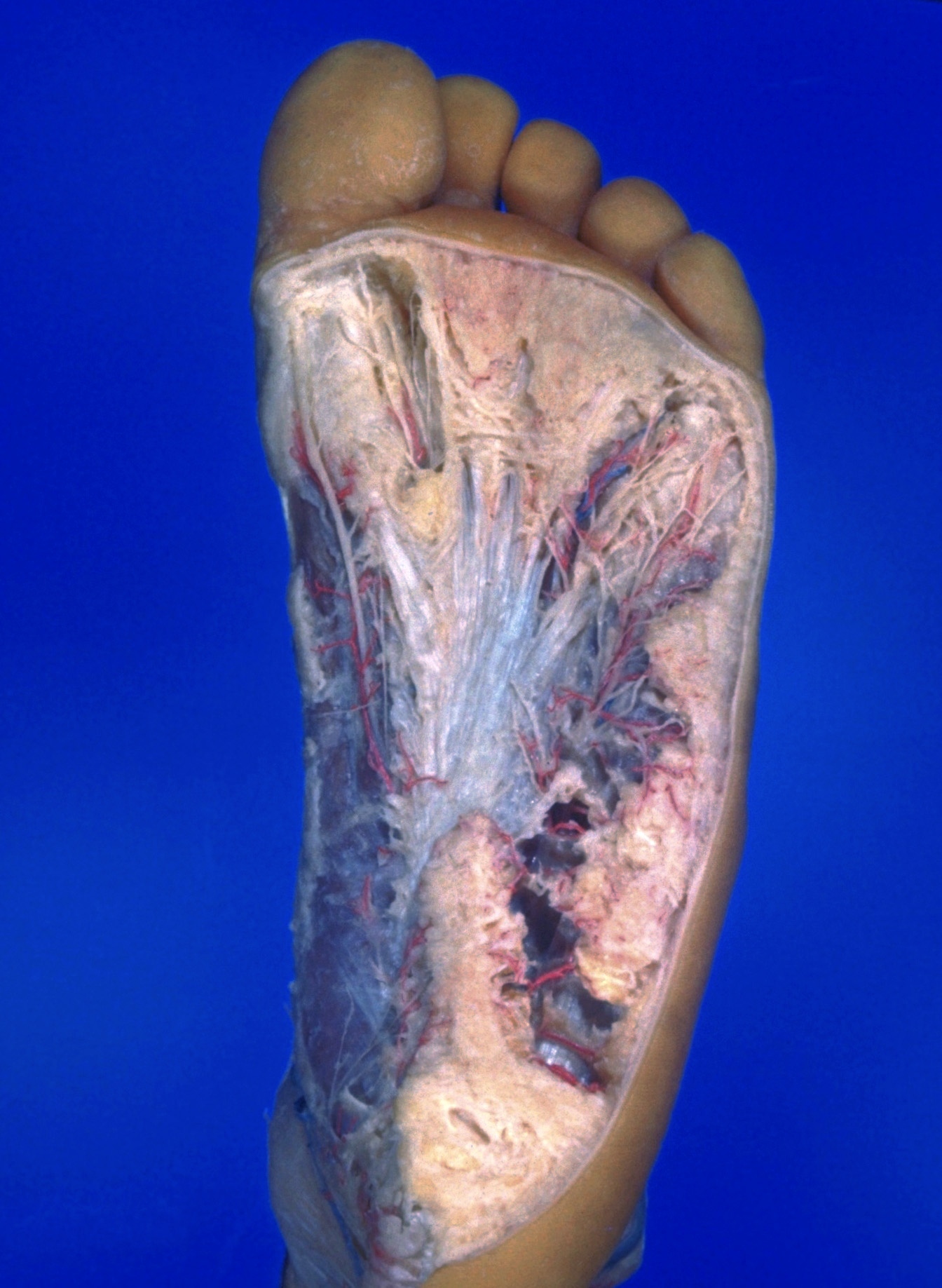
Dissection of plantar aspect of left foot

Tela subcutanea; superficial vessels and nerves of plantar surface of foot

Stanford holds the copyright to the David L. Bassett anatomical images and has assigned Creative Commons license Attribution-Share Alike 4.0 International to all of the images.
For additional information regarding use and permissions, please contact Dr. Drew Bourn at dbourn@stanford.edu.

Image #199-6
Bassett ImageDiagram Image
Legend Image
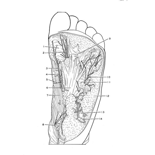
Dissection of plantar aspect of left foot
Tela subcutanea; superficial vessels and nerves of plantar surface of foot
A margin of skin has been preserved laterally and distally on the foot. The tela subcutanea has been variously dissected. In some areas it has been kept intact to illustrate the relative thickness of this layer on different parts of the foot. Elsewhere the superficial vessels and nerves and the plantar aponeurosis have been exposed.
1 . Fibrous sheath
2 . Proper plantar digital nerve
3 . Proper plantar digital nerve
4 . Branch of medial plantar artery
5 . Saphenous nerve (also see view 198-5)
6 . Plantar aponeurosis
7 . Abductor hallucis muscle (covered by thin medial part of plantar aponeurosis)
8 . Calcaneal branch of medial tibial nerve
9 . Transverse fasciculus of plantar aponeurosis
10 . Branch of lateral plantar nerve
11 . Branch of lateral plantar artery
12 . Tela subcutanea
13 . Abductor digiti minimi muscle (covered by lateral portion of plantar aponeurosis)
14 . Branch of lateral plantar artery