X CLOSE

Dissection of dorsolateral aspect of left foot and ankle

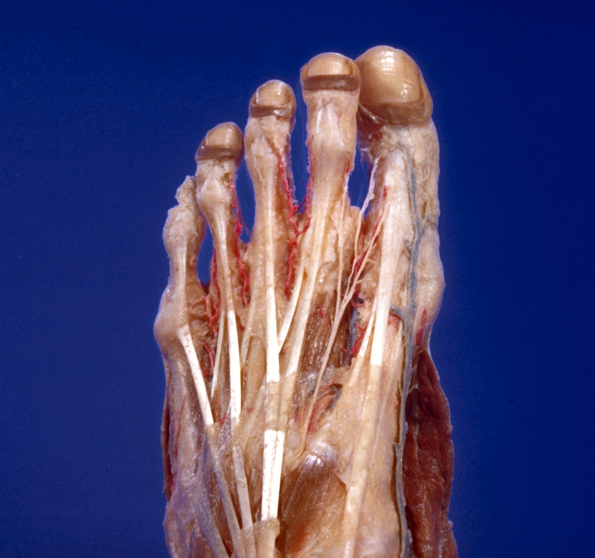
Blood vessels and extensor tendons of toes; terminal branches of deep peroneal nerve, viewed from above

Stanford holds the copyright to the David L. Bassett anatomical images and has assigned Creative Commons license Attribution-Share Alike 4.0 International to all of the images.
For additional information regarding use and permissions, please contact Dr. Drew Bourn at dbourn@stanford.edu.

Image #198-2
Bassett ImageDiagram Image
Legend Image
Dissection of dorsolateral aspect of left foot and ankle
Blood vessels and extensor tendons of toes; terminal branches of deep peroneal nerve, viewed from above
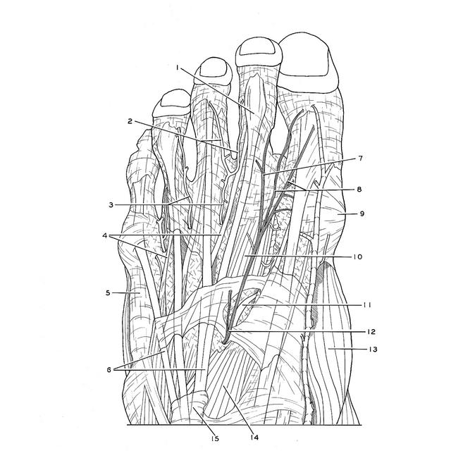
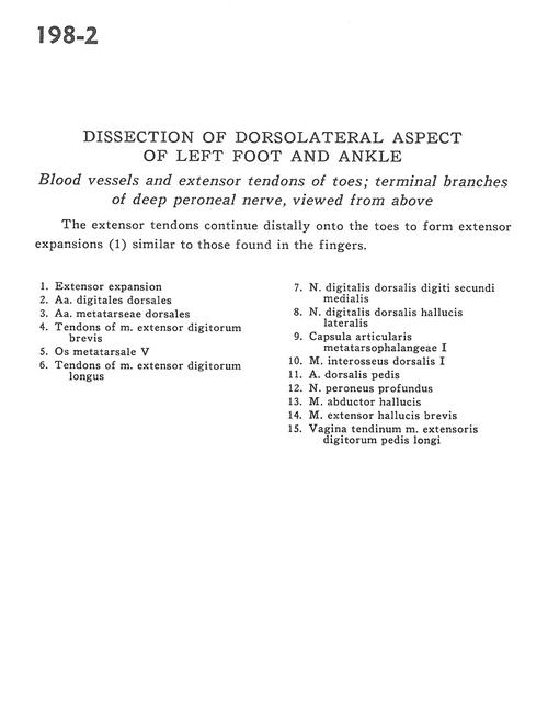
The extensor tendons continue distally onto the toes to form extensor expansions (1) similar to those found in the fingers.
1 . Extensor expansion
2 . Dorsal digital arteries
3 . Dorsal metatarsal arteries
4 . Tendons of extensor digitorum brevis muscle
5 . 5th metatarsal bone
6 . Tendons of extensor digitorum longus muscle
7 . Dorsal digital nerve to medial second toe
8 . Dorsal digital nerve to lateral first toe
9 . Capsule of 1st metatarsophalangeal joint
10 . 1st dorsal interosseus muscle
11 . Dorsalis pedis artery
12 . Deep peroneal nerve
13 . Abductor hallucis muscle
14 . Extensor hallucis brevis muscle
15 . Sheath of extensor digitorum longus tendon