Dissection of dorsolateral aspect of left foot and ankle
Nerve supply to abductor digiti minimi muscle, close-up lateral view
Stanford holds the copyright to the David L. Bassett anatomical images and has assigned
Creative Commons license Attribution-Share
Alike 4.0 International to all of the images.
For additional information regarding use and permissions,
please contact Dr. Drew Bourn at dbourn@stanford.edu.
Image #197-6
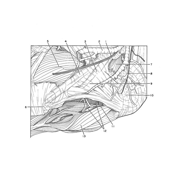
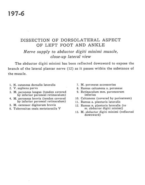
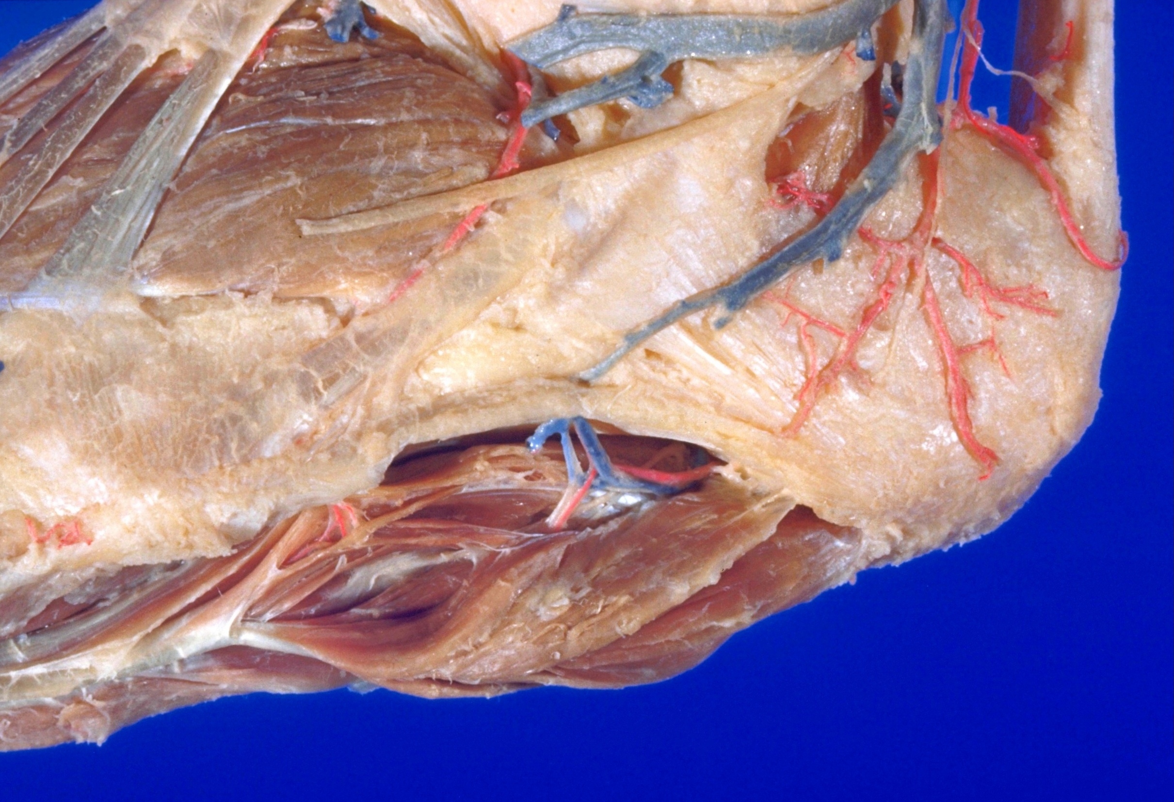
Dissection of dorsolateral aspect of left foot and ankle
Nerve supply to abductor digiti minimi muscle, close-up lateral view
The abductor digiti minimi has been reflected downward to expose the branch of the lateral plantar nerve (12) as it passes within the substance of the muscle.
- Dorsal lateral cutaneous nerve
- Greater saphenous vein
- Peroneus longus muscle (tendon covered by inferior peroneal retinaculum)
- Peroneus brevis muscle (tendon covered by inferior peroneal retinaculum)
- Extensor digitorum brevis muscle
- Tuberosity of 5th metatarsal bone
- Accessory peroneus muscle
- Calcaneal branch of peroneal artery
- Inferior peroneal retinaculum
- Calcaneus (covered by periosteum)
- Branch of lateral plantar artery
- Branch of lateral plantar nerve (to abductor digiti minimi muscle)
- Abductor digiti minimi muscle (reflected downward)