Exploration of those parts of the brain supplied by the posterior cerebral artery
Cingulum and inferior longitudinal fasciculus
Stanford holds the copyright to the David L. Bassett anatomical images and has assigned
Creative Commons license Attribution-Share
Alike 4.0 International to all of the images.
For additional information regarding use and permissions,
please contact Dr. Drew Bourn at dbourn@stanford.edu.
Image #19-3
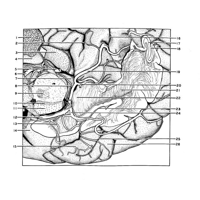
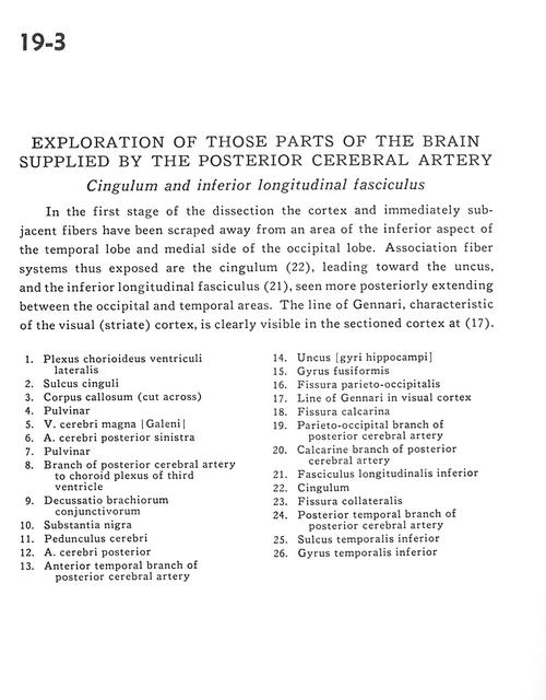
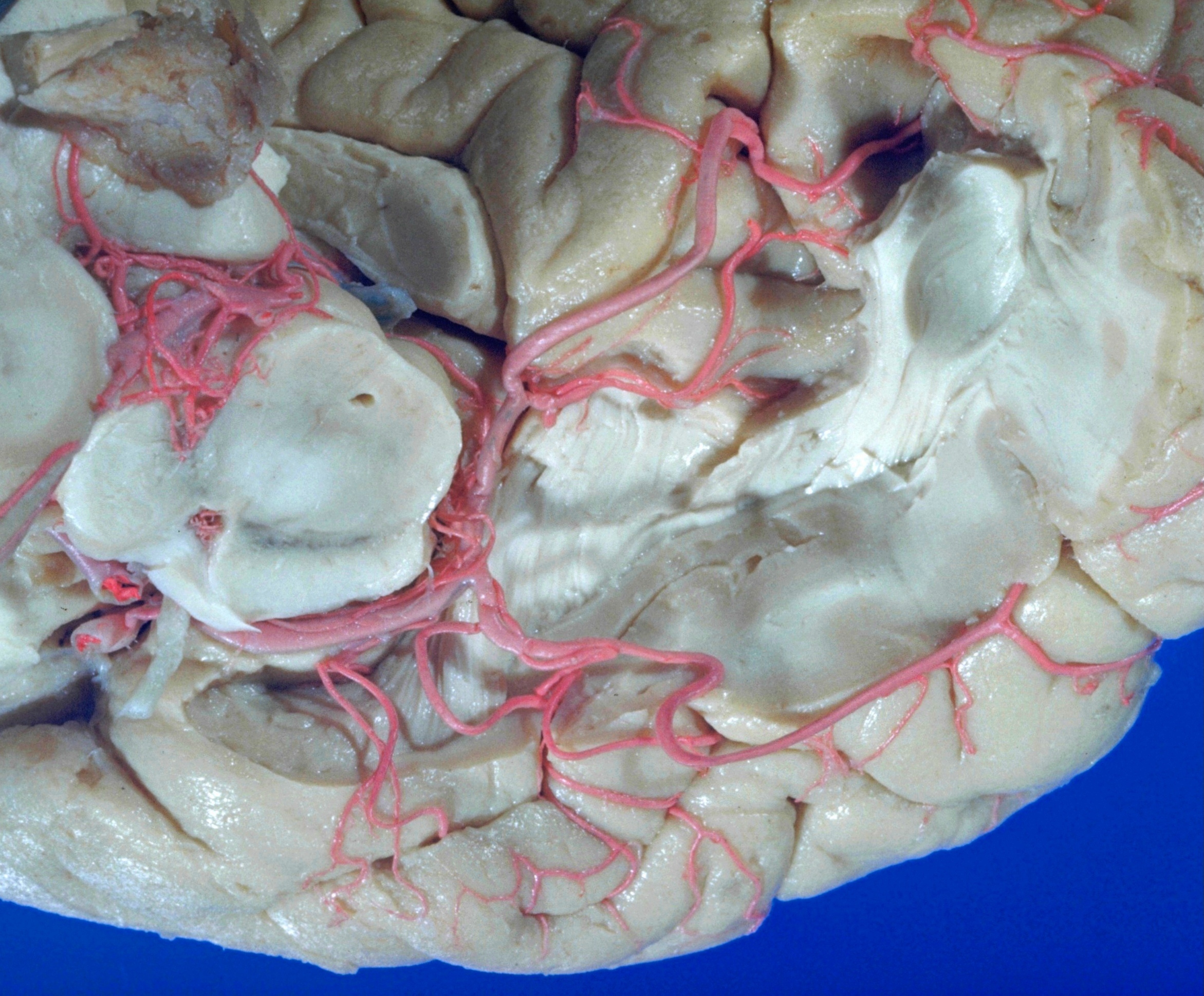
Exploration of those parts of the brain supplied by the posterior cerebral artery
Cingulum and inferior longitudinal fasciculus
In the first stage of the dissection the cortex and immediately subjacent fibers have been scraped away from an area of the inferior aspect of the temporal lobe and medial side of the occipital lobe. Association fiber systems thus exposed are the cingulum (22), leading toward the uncus, and the inferior longitudinal fasciculus (22), seen more posteriorly extending between the occipital and temporal areas. The line of Gennari, characteristic of the visual (striate) cortex, is clearly visible in the sectioned cortex at (17).
- Choroid plexus of lateral ventricle
- Cingulate sulcus
- Corpus callosum (cut across)
- Pulvinar
- Great cerebral vein (of Galen)
- Left posterior cerebral artery
- Pulvinar
- Branch of posterior cerebral artery to choroid plexus of third ventricle
- Decussation of brachium conjunctivum
- Substantia nigra
- Cerebral peduncles
- Posterior cerebral artery
- Anterior temporal branch of posterior cerebral artery
- Uncus (hippocampal gyri)
- Fusiform gyrus
- Parieto-occipital fissure
- Line of Gennari in visual cortex
- Calcarine fissure
- Parieto-occipital branch of posterior cerebral arteiy
- Calcarine branch of posterior cerebral artery
- Inferior longitudinal fasciculus
- Cingulum
- Collateral fissure
- Posterior temporal branch of posterior cerebral artery
- Inferior temporalis sulcus
- Inferior temporal gyrus.