X CLOSE

Dissection of knee

Superficial vessels and nerves of left knee, anterior view

Stanford holds the copyright to the David L. Bassett anatomical images and has assigned Creative Commons license Attribution-Share Alike 4.0 International to all of the images.
For additional information regarding use and permissions, please contact Dr. Drew Bourn at dbourn@stanford.edu.

Image #189-4
Bassett ImageDiagram Image
Legend Image
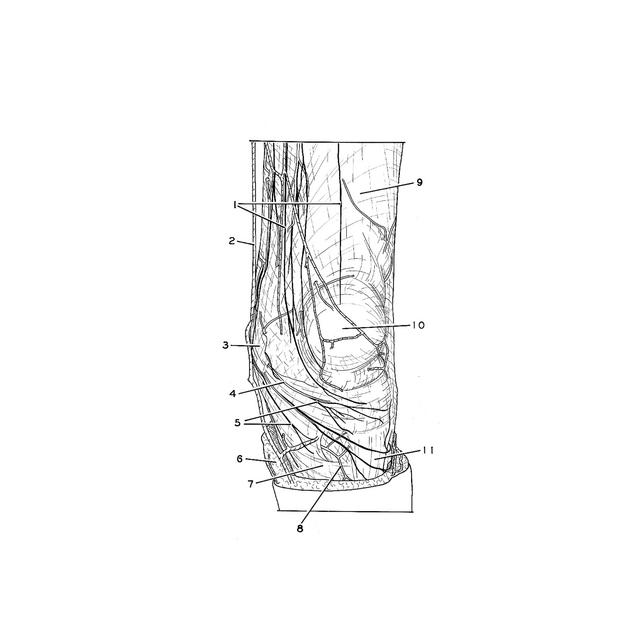
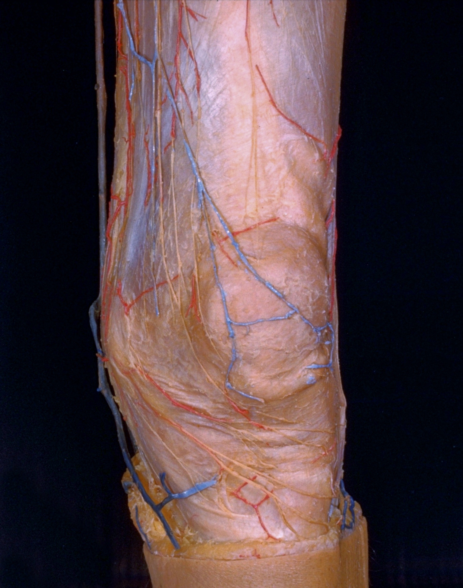
Dissection of knee
Superficial vessels and nerves of left knee, anterior view
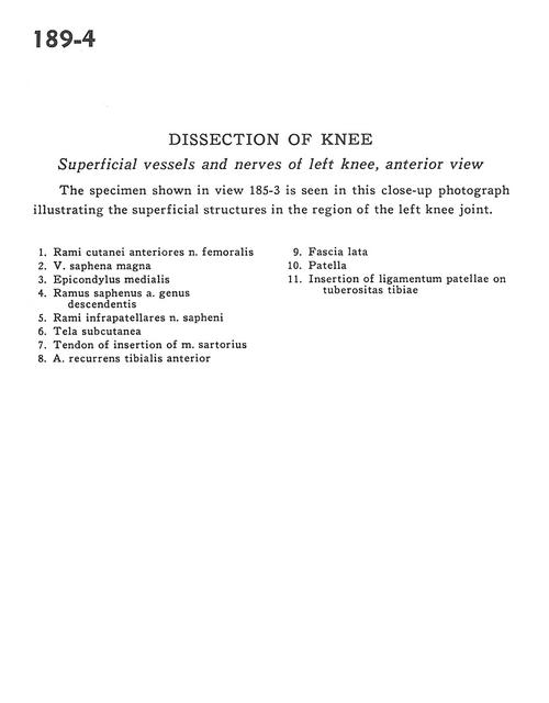
The specimen shown in view 185-3 is seen in this close-up photograph illustrating the superficial structures in the region of the left knee joint.
1 . Cutaneous branches of anterior femoral nerve
2 . Greater saphenous vein
3 . Medial epicondyle
4 . Saphenous branch of descending genicular artery
5 . Infrapatellar branch of saphenous nerve
6 . Tela subcutanea
7 . Tendon of insertion of sartorius muscle
8 . Anterior tibial recurrent artery
9 . Fascia lata
10 . Patella
11 . Insertion of patellar ligament on tibial tuberosity