Dissection of anterior and medial aspects of thigh
Nerve supply to rectus femoris muscle
Stanford holds the copyright to the David L. Bassett anatomical images and has assigned
Creative Commons license Attribution-Share
Alike 4.0 International to all of the images.
For additional information regarding use and permissions,
please contact Dr. Drew Bourn at dbourn@stanford.edu.
Image #187-2
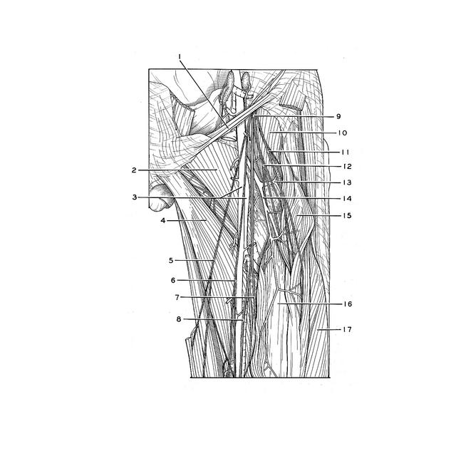
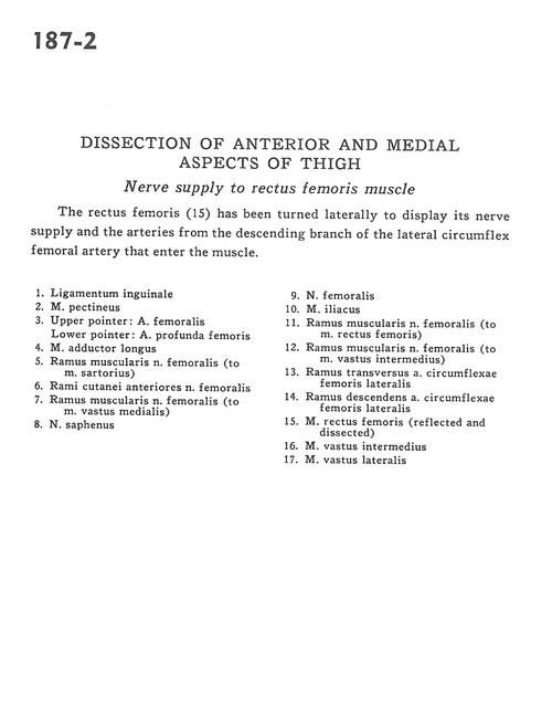
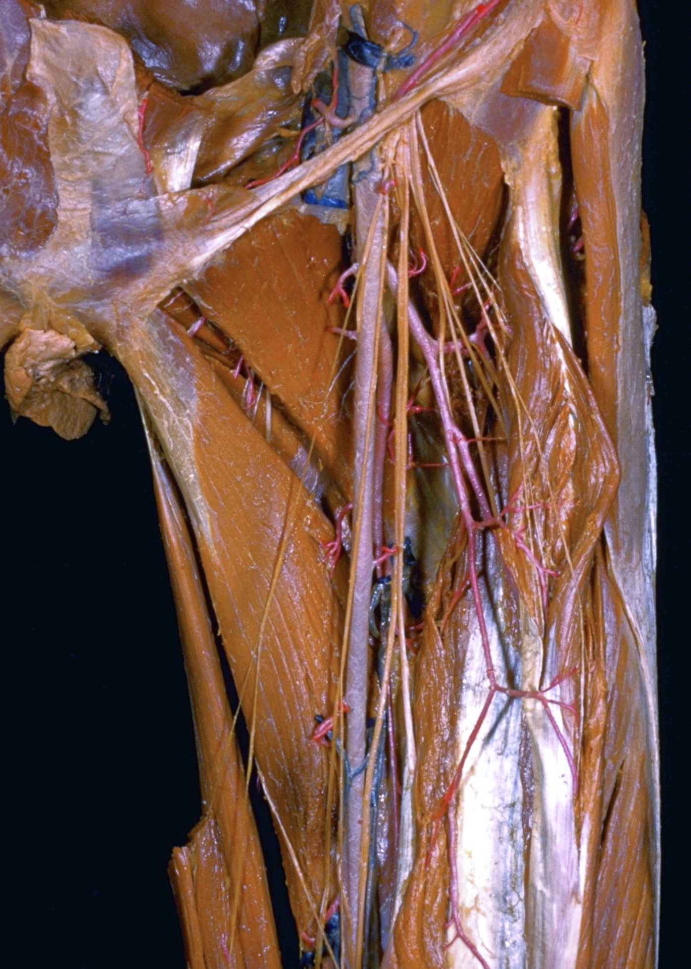
Dissection of anterior and medial aspects of thigh
Nerve supply to rectus femoris muscle
The rectus femoris (15) has been turned laterally to display its nerve supply and the arteries from the descending branch of the lateral circumflex femoral artery that enter the muscle.
- Inguinal ligament
- Pectineus muscle
- Upper pointer: Femoral artery Lower pointer: Deep femoral artery
- Adductor longus muscle
- Muscular branch of femoral nerve (to sartorius muscle)
- Cutaneous branches of anterior femoral nerve
- Muscular branch of femoral nerve (to vastus medialis muscle)
- Saphenous nerve
- Femoral nerve
- Iliacus muscle
- Muscular branch of femoral nerve (to rectus femoris muscle)
- Muscular branch of femoral nerve (to vastus intermedius muscle)
- Transverse branch of lateral femoral circumflex artery
- Descending branch of lateral femoral circumflex artery
- Rectus femoris muscle (reflected and dissected)
- Vastus intermedius muscle
- Vastus lateralis muscle