Dissection of anterior and medial aspects of thigh
Nerve supply to sartorius and pectineus muscles; branches of femoral artery
Stanford holds the copyright to the David L. Bassett anatomical images and has assigned
Creative Commons license Attribution-Share
Alike 4.0 International to all of the images.
For additional information regarding use and permissions,
please contact Dr. Drew Bourn at dbourn@stanford.edu.
Image #186-7
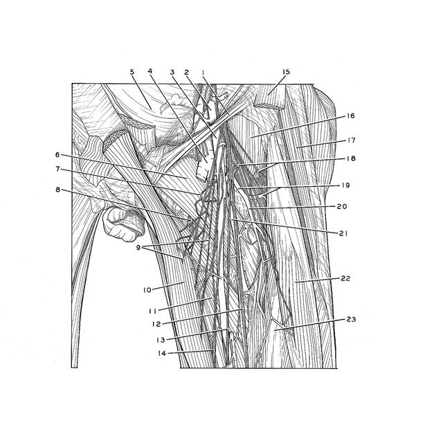
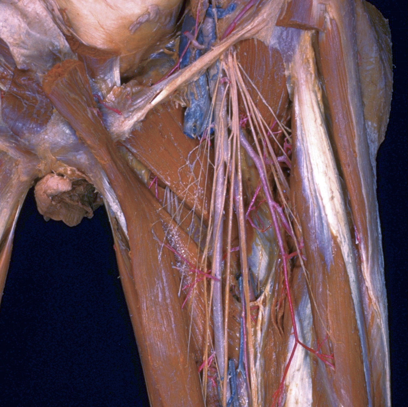
Dissection of anterior and medial aspects of thigh
Nerve supply to sartorius and pectineus muscles; branches of femoral artery
The sartorius muscle has been divided close to its origin. The muscle belly has been pulled medially toward the pubis to reveal the course of branches of the femoral nerve into the upper part of the muscle. View 187-4 illustrates the nerves in the lower part of the sartorius. The nerve to the pectineus (7) passes medially behind the femoral vessels to enter the muscle. In this dissection the femoral vein and most of its branches within the femoral triangle have been resected.
- Inguinal ligament
- External iliac artery
- Femoral artery
- Femoral vein (cut off)
- Peritoneum
- Pectineus muscle
- Muscular branch of femoral nerve (to Pectineus muscle)
- Anterior branch of obturator nerve (descending in front of adductor brevis)
- Muscular branch femoral nerve (to sartorius muscle)
- Sartorius muscle (reflected; also see no. 15)
- Adductor longus muscle
- Muscular branch of femoral nerve (to Vastus medialis muscle)
- Saphenous nerve
- Muscular branch of femoral nerve (to sartorius muscle)
- Sartorius muscle (origin)
- Iliacus muscle
- Tensor fasciae latae muscle
- Muscular branch femoral nerve
- Upper pointer: Lateral femoral circumflex artery Lower pointer: Transverse branch of lateral femoral circumflex artery
- Descending branch of lateral femoral circumflex artery
- Deep femoral artery
- Rectus femoris muscle (reflected)
- Vastus intermedius muscle