X CLOSE

Exploration of gluteal region and hip

General view of gluteal muscles

Stanford holds the copyright to the David L. Bassett anatomical images and has assigned Creative Commons license Attribution-Share Alike 4.0 International to all of the images.
For additional information regarding use and permissions, please contact Dr. Drew Bourn at dbourn@stanford.edu.

Image #179-3
Bassett ImageDiagram Image
Legend Image
Exploration of gluteal region and hip
General view of gluteal muscles
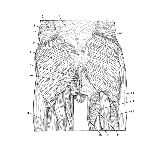
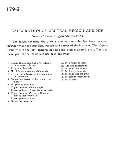
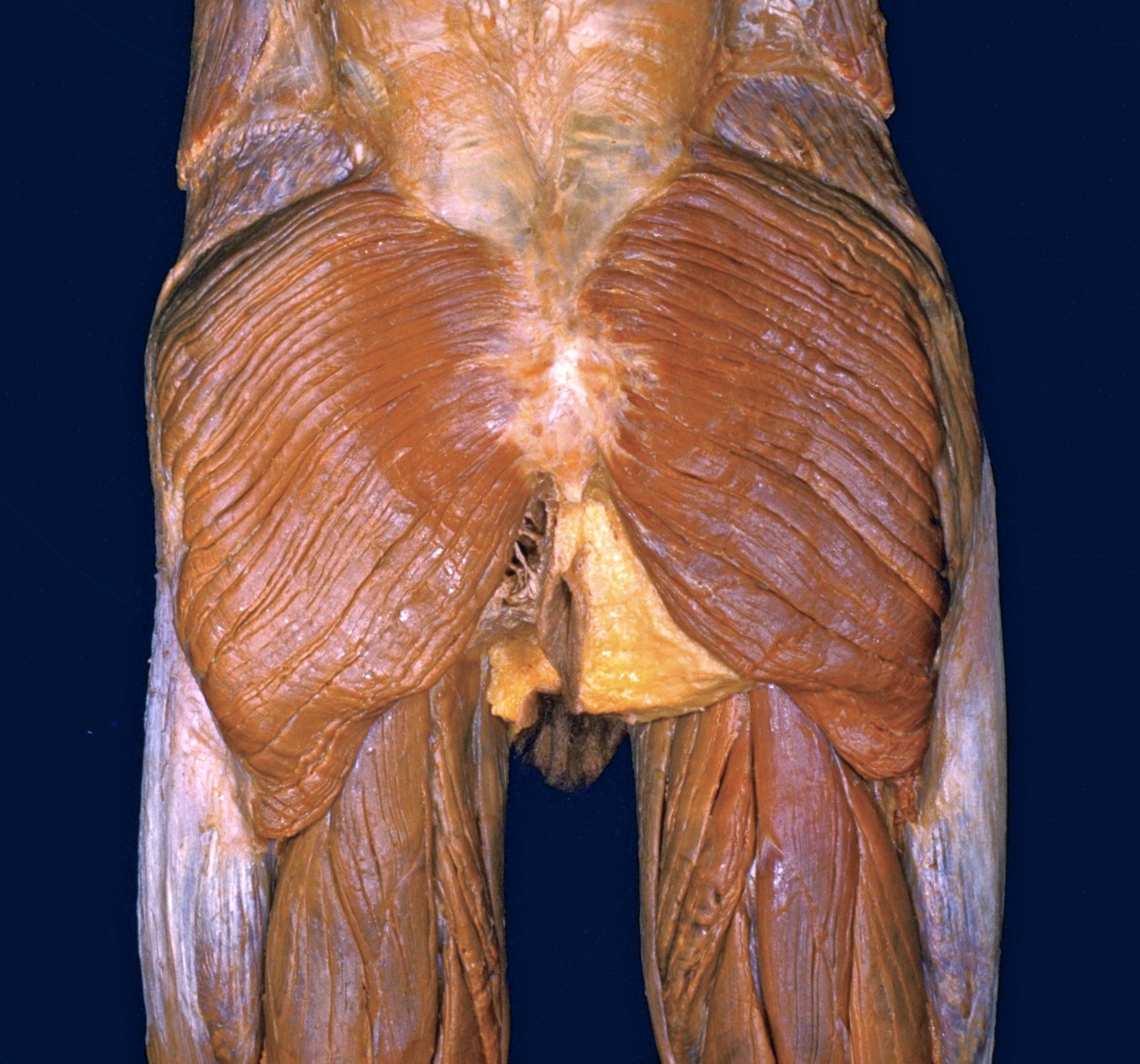
The fascia covering the gluteus maximus muscles has been resected, together with the superficial vessels and nerves of the buttocks. The adipose tissue within the left ischiorectal fossa has been dissected away. The posterior part of the fascia lata has been cut away.
1 . Thoracolumbar fascia (overlying erector spinae muscle)
2 . Lumbar triangle
3 . External oblique muscle
4 . Iliac crest (covered by fascia and periosteum)
5 . Sacral bone (covered by connective tissue)
6 . Gluteus maximus muscle
7 . Upper pointer: Coccyx bone Lower pointer: Ischiorectal fossa
8 . Upper pointer: Adipose body of ischiorectal fossa Lower pointer: Anus
9 . Vastus lateralis muscle
10 . Gluteus medius muscle
11 . Iliotibial tract
12 . Semitendinosus muscle
13 . Biceps femoris muscle
14 . Adductor magnus muscle
15 . Semimembranosus muscle
16 . Gracilis muscle