Exploration of the brain from the medial aspect
Optic tract, internal capsule, medial surface of temporal lobe and posterior cerebral artery
Stanford holds the copyright to the David L. Bassett anatomical images and has assigned
Creative Commons license Attribution-Share
Alike 4.0 International to all of the images.
For additional information regarding use and permissions,
please contact Dr. Drew Bourn at dbourn@stanford.edu.
Image #17-6
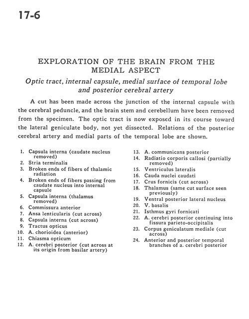
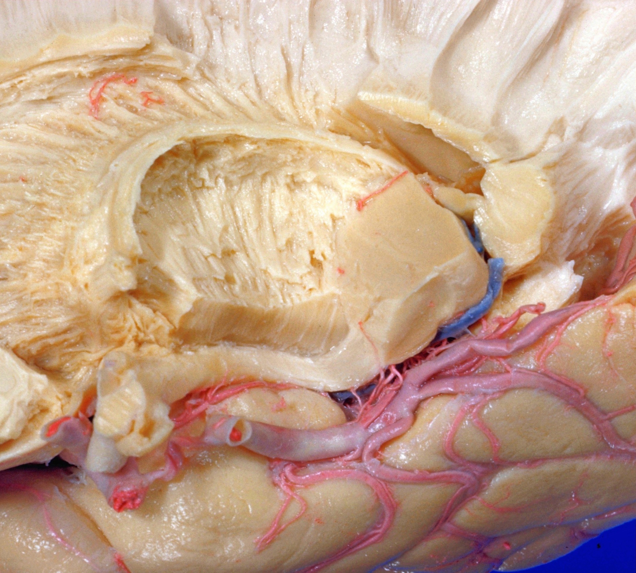
Exploration of the brain from the medial aspect
Optic tract, internal capsule, medial surface of temporal lobe and posterior cerebral artery
A cut has been made across the junction of the internal capsule with the cerebral peduncle, and the brain stem and cerebellum have been removed from the specimen. The optic tract is now exposed in its course toward the lateral geniculate body, not yet dissectd. Relations of the posterior cerebral artery and medial parts of the temporal lobe are shown.
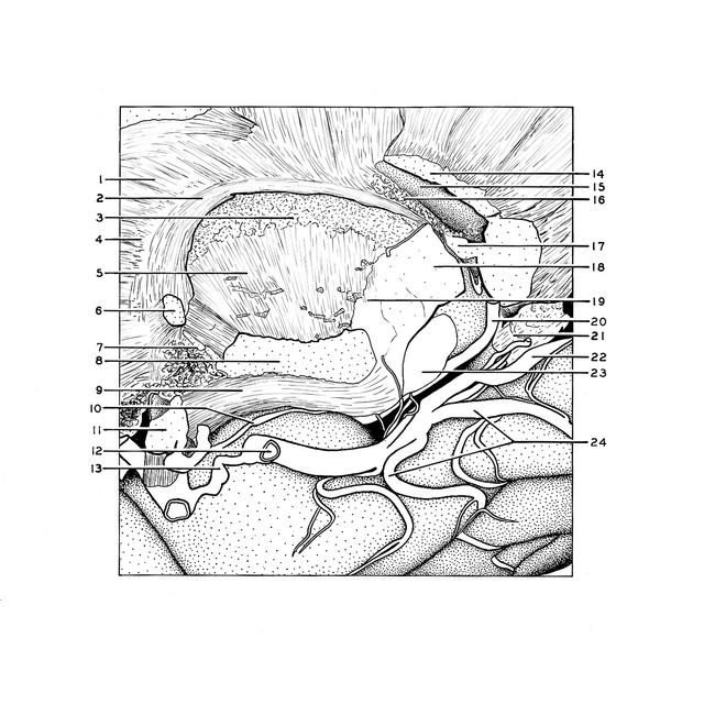
- Internal capsule (caudate nucleus removed)
- Stria terminalis
- Broken ends of fibers of thalamic radiation
- Broken ends of fibers passing from caudate nucleus into internal capsule
- Internal capsule (thalamus removed)
- Anterior commissure
- Ansa lenticularis (cut across)
- Internal capsule (cut across)
- Optic tract
- Choroidal artery (anterior)
- Optic chiasm
- Posterior cerebral artery (cut across at its origin from basilar artery)
- Posterior communicating artery
- Radiation corpus callosum (partially removed)
- Lateral ventricle
- Caudate nucleus (tail)
- Fornix (crus) (cut across)
- Thalamus (same cut surface seen previously)
- Ventral posterior lateral nucleus
- Basal vein
- Limbic lobe
- Posterior cerebral artery continuing into parietooccipital fissure
- Medial geniculate body (cut across)
- Anterior and posterior temporal branches of posterior cerebral artery