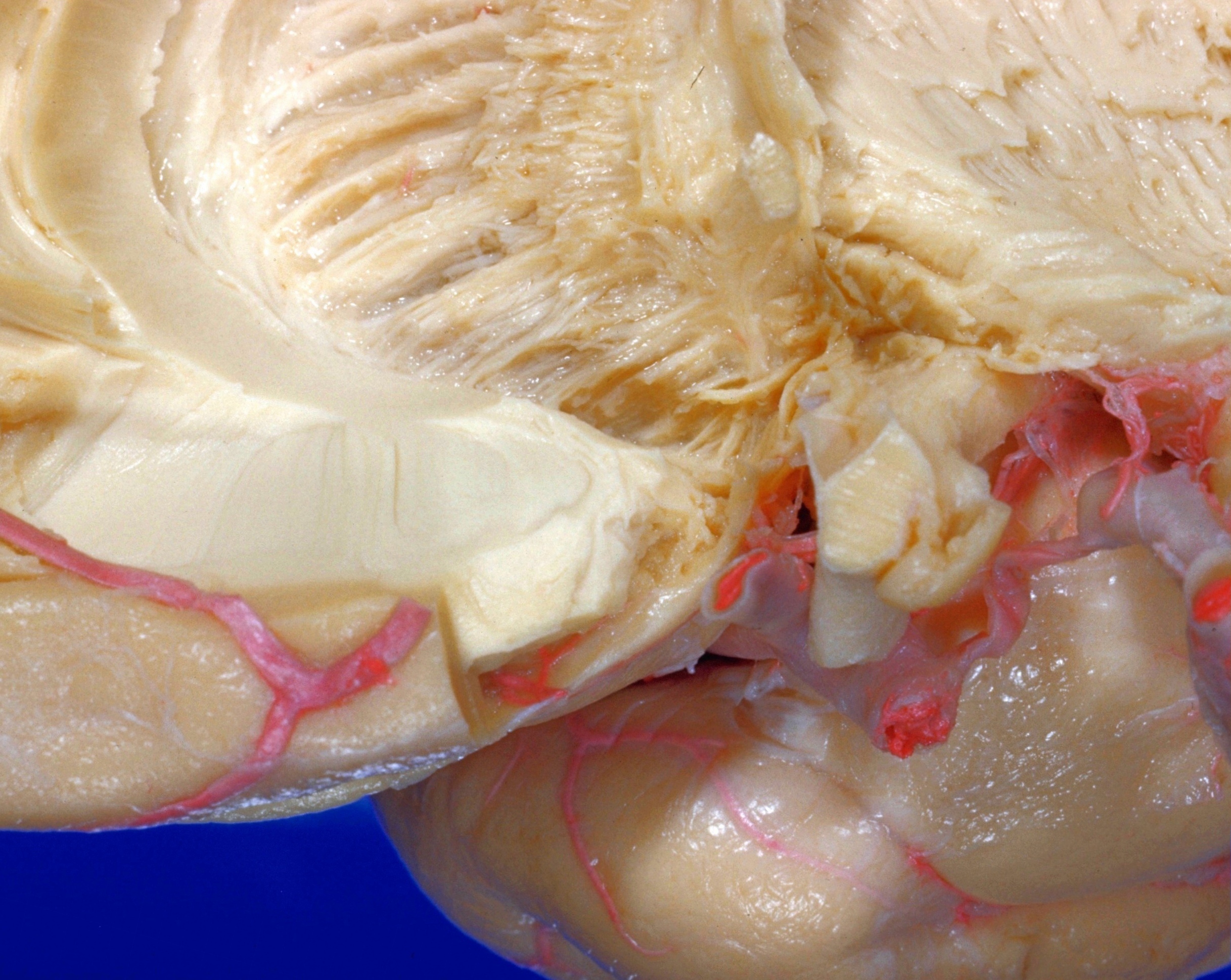
Exploration of the brain from the medial aspect
Anterior limb of anterior commissure
Stanford holds the copyright to the David L. Bassett anatomical images and has assigned
Creative Commons license Attribution-Share
Alike 4.0 International to all of the images.
For additional information regarding use and permissions,
please contact Dr. Drew Bourn at dbourn@stanford.edu.
Image #17-2
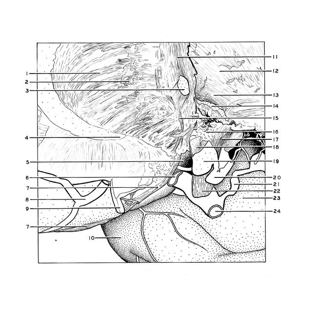
Exploration of the brain from the medial aspect
Anterior limb of anterior commissure
The anterior limb (15) of the anterior commissure is seen in this view as it diverges from the posterior limb and approaches the olfactory trigone (5). The gyrus rectus (9) has been cut away to expose the olfactory tract (7) in its normal position within the olfactory sulcus. The cut end of the anterior commissure is in the midline.
- Internal capsule
- Broken ends of fibers from head of caudate nucleus into internal capsule
- Anterior commissure
- Cut surface of radiation of corpus callosum
- Olfactory trigone
- Anterior cerebral artery (divided)
- Olfactory tract
- Orbital branch of anterior cerebral artery
- Straight gyrus (cortex removed posteriorly)
- Inferior temporal gyrus
- Stria terminalis
- Internal capsule
- Inferior margin of internal capsule
- Ansa lenticularis (partially cut away)
- Anterior part of anterior commissure
- Lateral region of hypothalamus
- Optic tract
- Optic recess (ependymal surface still present)
- Optic chiasm
- Infundibulum
- Posterior communicating artery
- Optic nerve (II)
- Uncus (hippocampal gyrus)
- Internal carotid artery