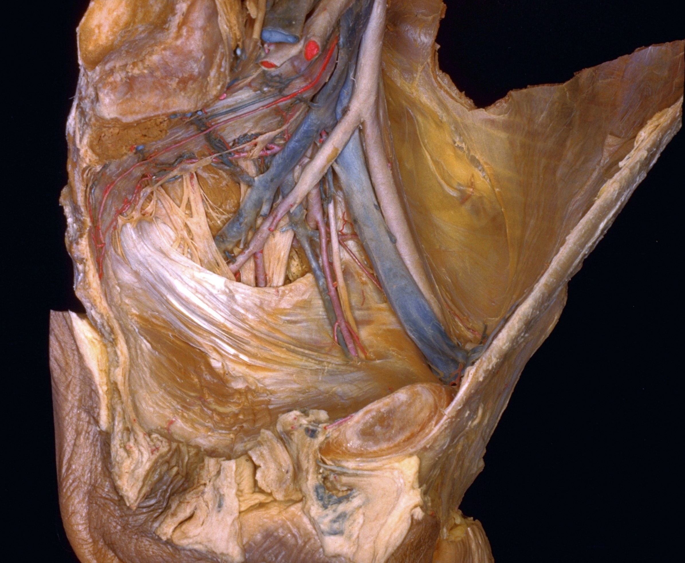
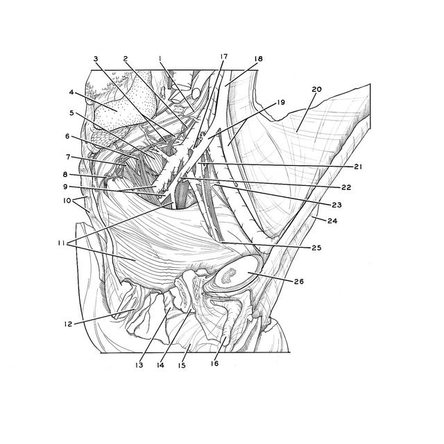
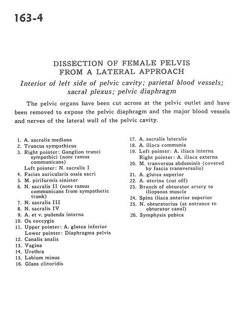
Dissection of female pelvis from a lateral approach
Interior of left side of pelvic cavity; parietal blood vessels; sacral plexus; pelvic diaphragm
Stanford holds the copyright to the David L. Bassett anatomical images and has assigned
Creative Commons license Attribution-Share
Alike 4.0 International to all of the images.
For additional information regarding use and permissions,
please contact Dr. Drew Bourn at dbourn@stanford.edu.
Image #163-4
Dissection of female pelvis from a lateral approach
Interior of left side of pelvic cavity; parietal blood vessels; sacral plexus; pelvic diaphragm
The pelvic organs have been cut across at the pelvic outlet and have been removed to expose the pelvic diaphragm and the major blood vessels and nerves of the lateral wall of the pelvic cavity.
- Middle sacral artery
- Sympathetic trunk
- Right pointer: Ganglion of sympathetic trunk (note ramus communicans) Left pointer: Sacral nerve
- Articular surface of sacrum
- Left piriform muscle
- Sacral nerve II (note ramus communicans from sympathetic trunk)
- Sacral nerve III
- Sacral nerve IV
- Internal pudendal artery and vein
- Coccyx
- Upper pointer: Inferior gluteal artery Lower pointer: Pelvic diaphragm
- Anal canal
- Vagina
- Urethra
- Labium minus
- Glans of clitoris
- Lateral sacral artery
- Common iliac artery
- Left pointer: Internal iliac artery Right pointer: External iliac artery
- Transversus abdominis muscle (covered by transversalis fascia)
- Superior gluteal artery
- Uterine artery (cut off)
- Branch of obturator artery to iliopsoas muscle
- Anterior superior iliac spine
- Obturator nerve (at entrance to obturator canal)
- Pubic symphysis