Dissection of female pelvis from a lateral approach
Interior of left side of pelvic cavity.
Stanford holds the copyright to the David L. Bassett anatomical images and has assigned
Creative Commons license Attribution-Share
Alike 4.0 International to all of the images.
For additional information regarding use and permissions,
please contact Dr. Drew Bourn at dbourn@stanford.edu.
Image #162-3
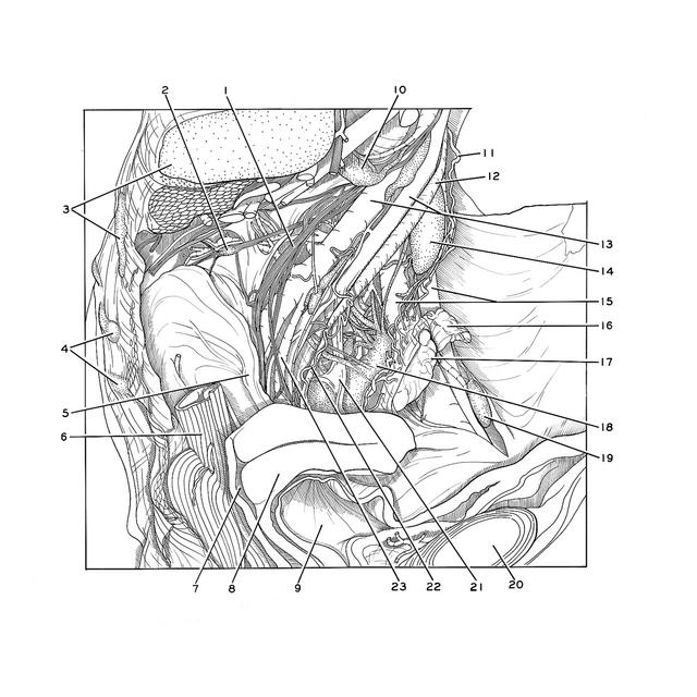
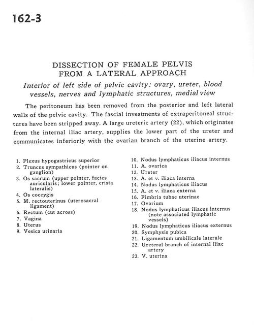
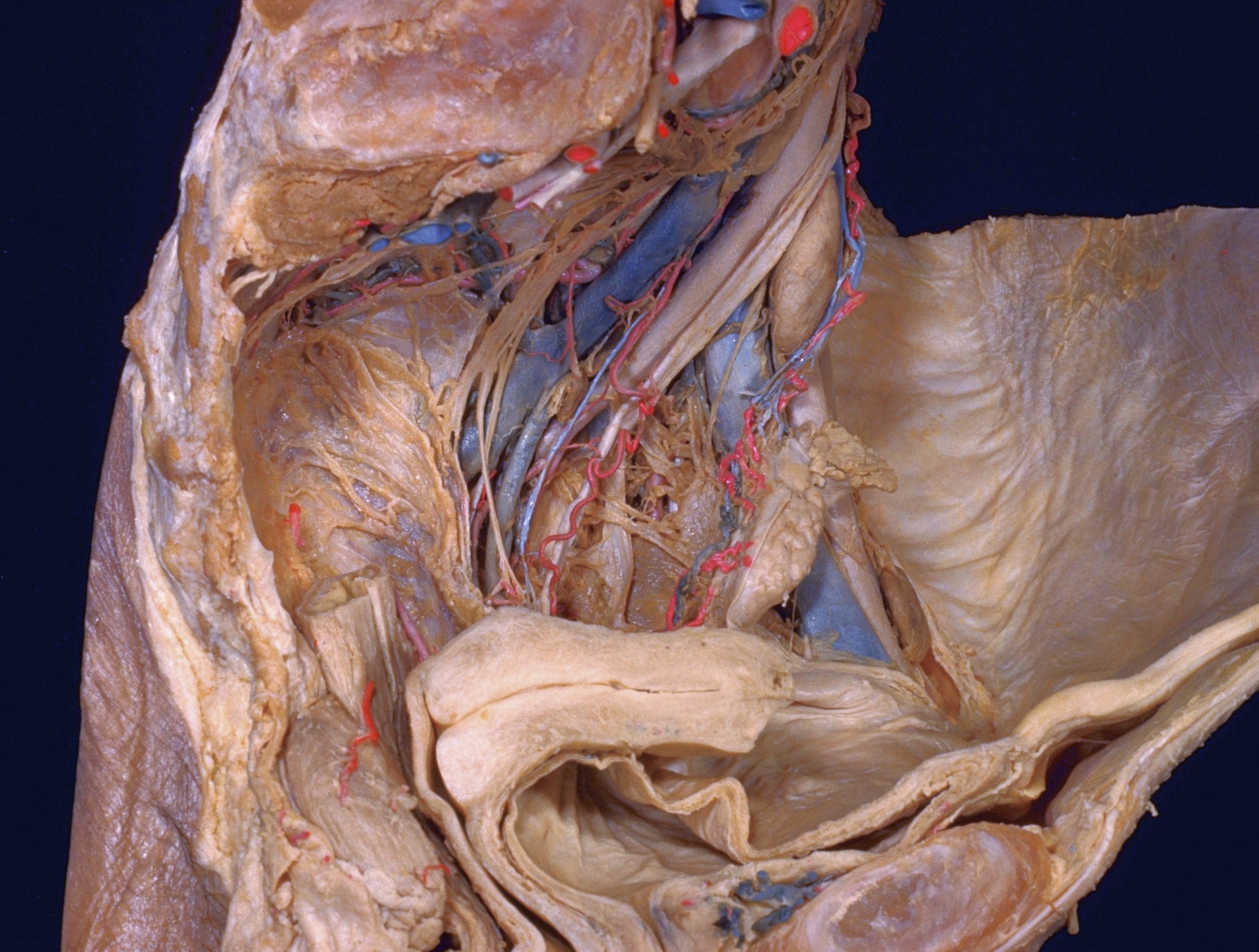
Dissection of female pelvis from a lateral approach
Interior of left side of pelvic cavity.
The peritoneum has been removed from the posterior and left lateral walls of the pelvic cavity. The fascial investments of extraperitoneal structures have been stripped away. A large ureteric artery (22), which originates from the internal iliac artery, supplies the lower part of the ureter and communicates inferiorly with the ovarian branch of the uterine artery.
- Superior hypogastric plexus
- Sympathetic trunk (pointer on ganglion)
- Sacrum (upper pointer, articular surface; lower pointer, lateral crest)
- Coccyx
- Rectouterinus muscle (uterosacral ligament)
- Rectum (cut across)
- Vagina
- Uterus
- Urinary bladder
- Internal iliac lymph node
- Ovarian artery
- Ureter
- Internal iliac artery and vein
- Iliac lymph node
- External iliac artery and vein
- Fimbria of uterine tube
- Ovary
- Internal iliac lymph node (note associated lymphatic vessels)
- External iliac lymph node
- Pubic symphysis
- Lateral umbilical ligament
- Ureteral branch of internal iliac artery
- Uterine vein