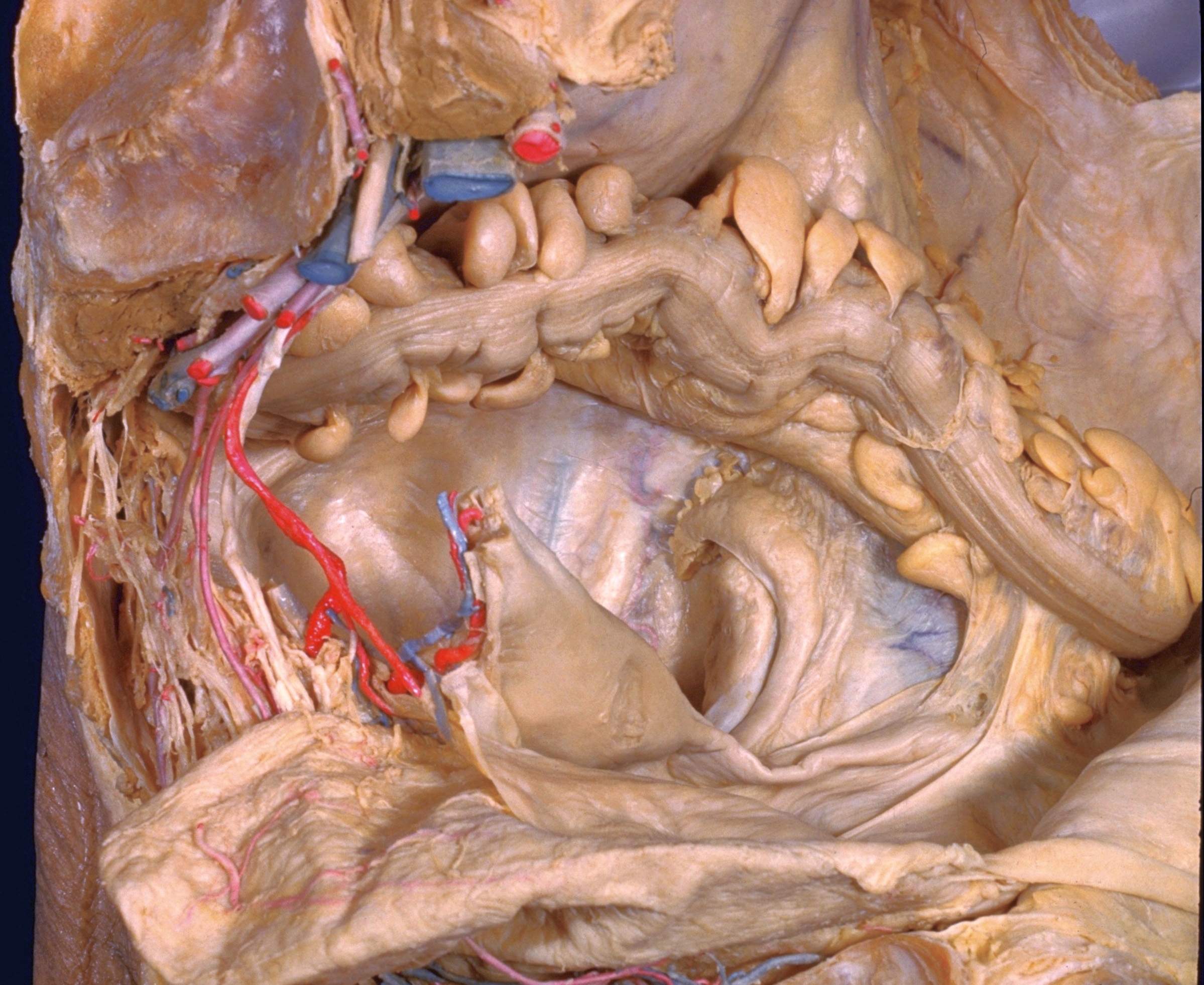
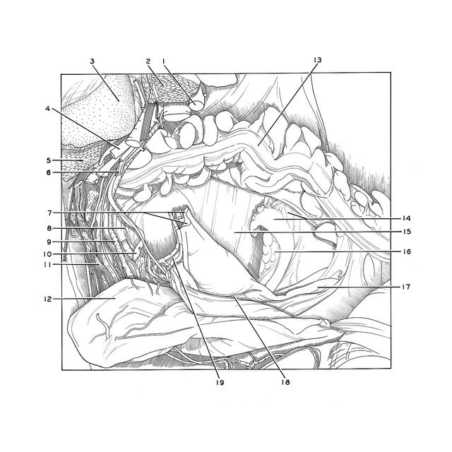
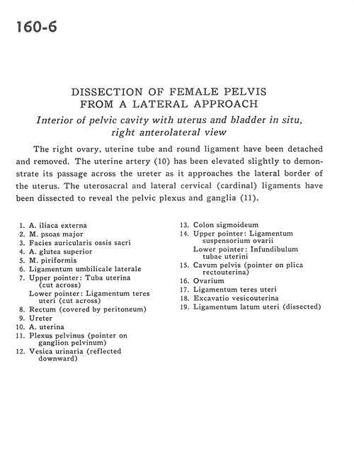
Dissection of female pelvis from a lateral approach
Interior of pelvic cavity with uterus and bladder in situ, right anterolateral view
Stanford holds the copyright to the David L. Bassett anatomical images and has assigned
Creative Commons license Attribution-Share
Alike 4.0 International to all of the images.
For additional information regarding use and permissions,
please contact Dr. Drew Bourn at dbourn@stanford.edu.
Image #160-6
Dissection of female pelvis from a lateral approach
Interior of pelvic cavity with uterus and bladder in situ, right anterolateral view
The right ovary, uterine tube and round ligament have been detached and removed. The uterine artery (10) has been elevated slightly to demonstrate its passage across the ureter as it approaches the lateral border of the uterus. The uterosacral and lateral cervical (cardinal) ligaments have been dissected to reveal the pelvic plexus and ganglia (11).
- External iliac artery
- Psoas major muscle
- Articular surface of sacrum
- Superior gluteal artery
- Piriform muscle
- Lateral umbilical ligament
- Upper pointer: Uterine tube (cut across) Lower pointer: Ligamentum teres (of uterus) (cut across)
- Rectum (covered by peritoneum)
- Ureter
- Uterine artery
- Pelvic plexus (pointer on pelvic ganglion)
- Urinary bladder (reflected downward)
- Sigmoid colon
- Upper pointer: Suspensory ligament of ovary Lower pointer: Infundibulum of uterine tube
- Pelvic cavity (pointer on rectouterine fold)
- Ovary
- Ligamentum teres (of uterus)
- Uterovesical pouch
- Broad ligament of uterus (dissected)