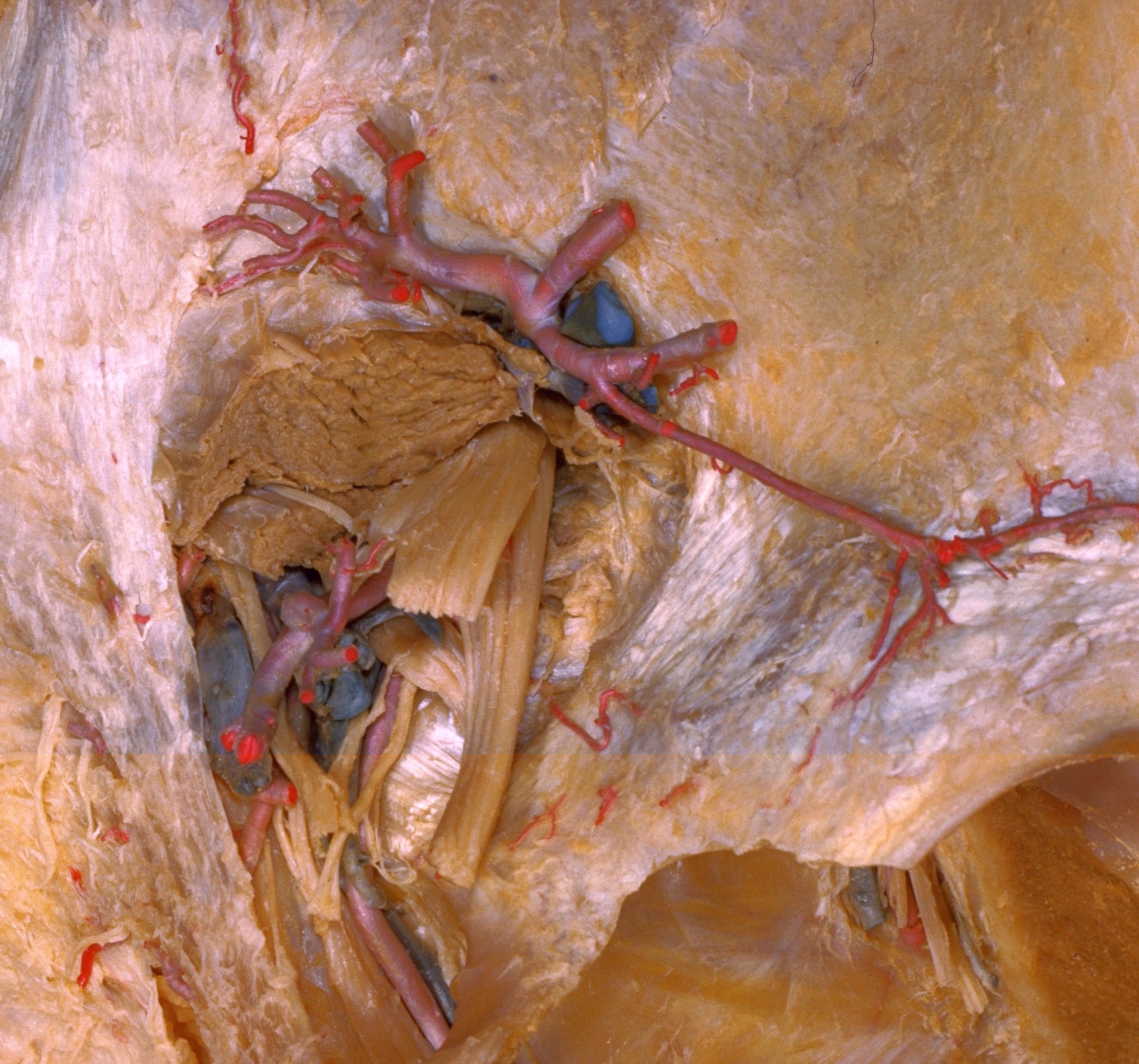
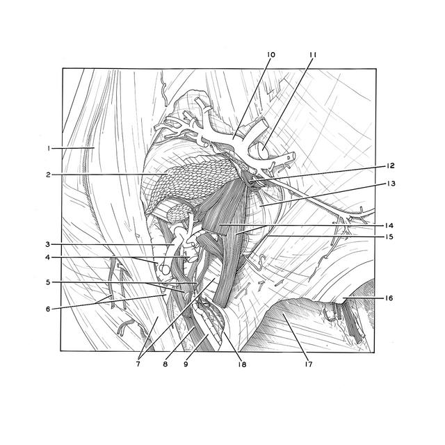
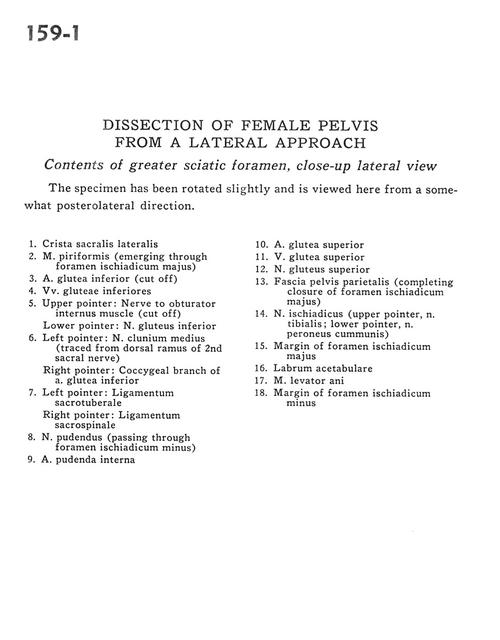
Dissection of female pelvis froma lateral approach
Contents of greater sciatic foramen, close-up lateral view
Stanford holds the copyright to the David L. Bassett anatomical images and has assigned
Creative Commons license Attribution-Share
Alike 4.0 International to all of the images.
For additional information regarding use and permissions,
please contact Dr. Drew Bourn at dbourn@stanford.edu.
Image #159-1
Dissection of female pelvis froma lateral approach
Contents of greater sciatic foramen, close-up lateral view
The specimen has been rotated slightly and is viewed here from a somewhat posterolateral direction.
- Lateral sacral crest
- Piriform muscle (emerging through greater sciatic foramen)
- Inferior gluteal artery (cut off)
- Inferior gluteal veins
- Upper pointer: Nerve to obturator internus muscle (cut off) Lower pointer: Inferior gluteal nerve
- Left pointer: Middle cluneal nerve (traced from dorsal ramus of 2nd sacral nerve) Right pointer: Coccygeal branch of inferior gluteal artery
- Left pointer: Sacrotuberous ligament Right pointer: Sacrospinous ligament
- Pudendal nerve (passing through lesser sciatic foramen)
- Internal pudendal artery
- Superior gluteal artery
- Superior gluteal vein
- Superior gluteal nerve
- Parietal pelvic fascia (completing closure of greater sciatic foramen)
- Sciatic nerve (upper pointer, tibial nerve; lower pointer, common peroneal nerve)
- Margin of greater sciatic foramen
- Acetabular labrum
- Levator ani muscle
- Margin of lesser sciatic foramen