Kidneys, suprarenal glands and posterior abdominal vessels, nerves and muscles
Nerves, blood vessels, and muscle attachments in relation to lumbar spine, left anterolateral view
Stanford holds the copyright to the David L. Bassett anatomical images and has assigned
Creative Commons license Attribution-Share
Alike 4.0 International to all of the images.
For additional information regarding use and permissions,
please contact Dr. Drew Bourn at dbourn@stanford.edu.
Image #151-7
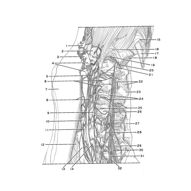
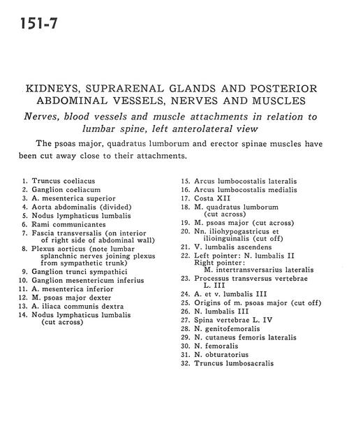
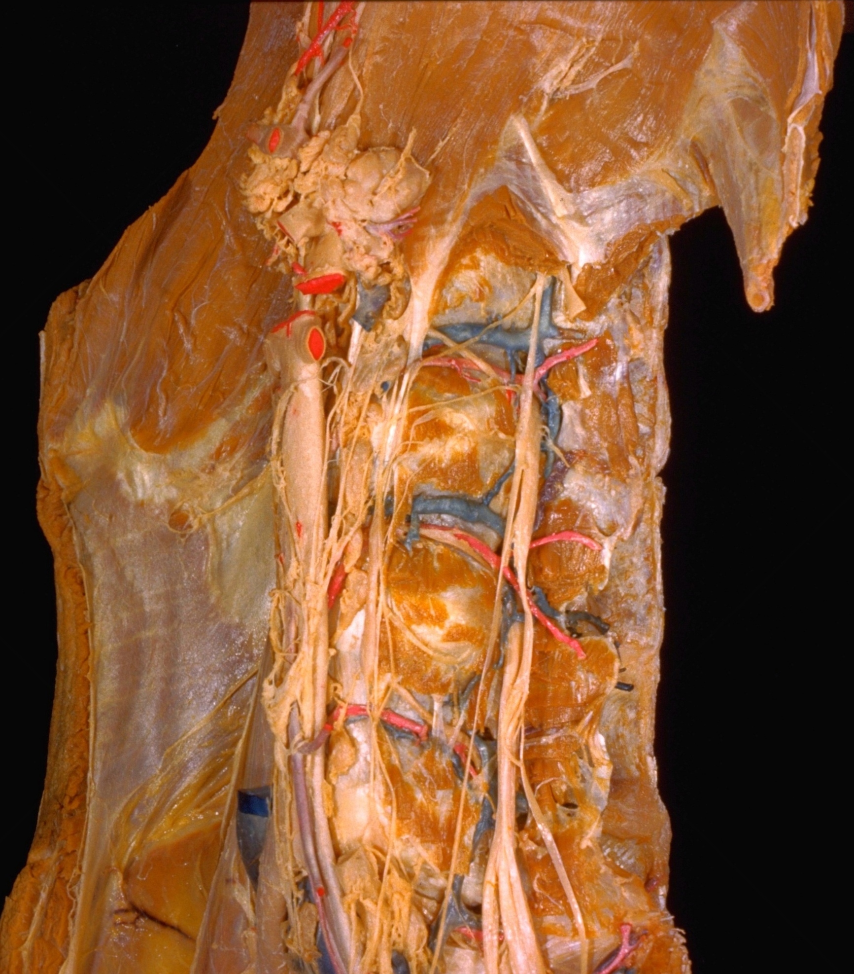
Kidneys, suprarenal glands and posterior abdominal vessels, nerves and muscles
Nerves, blood vessels, and muscle attachments in relation to lumbar spine, left anterolateral view
The psoas major, quadratus lumborum and erector spinae muscles have been cut away close to their attachments.
- Celiac trunk
- Celiac ganglion
- Superior mesenteric artery
- Abdominal aorta (divided)
- Lumbar lymph node
- Rami communicantes
- Transversalis fascia (on interior of right side of abdominal wall)
- Aortic plexus (note lumbar splanchnic nerves joining plexus from sympathetic trunk)
- Ganglion of sympathetic trunk
- Inferior mesenteric ganglion
- Inferior mesenteric artery
- Right psoas major muscle
- Right common iliac artery
- Lumbar lymph node (cut across)
- Lateral lumbocostal arch
- Medial lumbocostal arch
- Rib XII
- Quadratus lumborum muscle(cut across)
- Psoas major muscle (cut across)
- Iliohypogastric and ilioinguinal nerves (cut off)
- Ascending lumbar vein
- Left pointer: Lumbar nerve II Right pointer: Lateral intertransverse muscle
- Transverse process vertebra L. III
- Lumbar artery and vein III
- Origins of psoas major muscle (cut off)
- Lumbar nerve III
- Vertebral spine L. IV
- Genitofemoral nerve
- Lateral femoral cutaneous nerve
- Femoral nerve
- Obturator nerve
- Lumbosacral trunk