Kidneys, suprarenal glands and posterior abdominal vessels, nerves and muscles
Left kidney sectioned in situ; left suprarenal gland dissected, close-up view
Stanford holds the copyright to the David L. Bassett anatomical images and has assigned
Creative Commons license Attribution-Share
Alike 4.0 International to all of the images.
For additional information regarding use and permissions,
please contact Dr. Drew Bourn at dbourn@stanford.edu.
Image #151-1
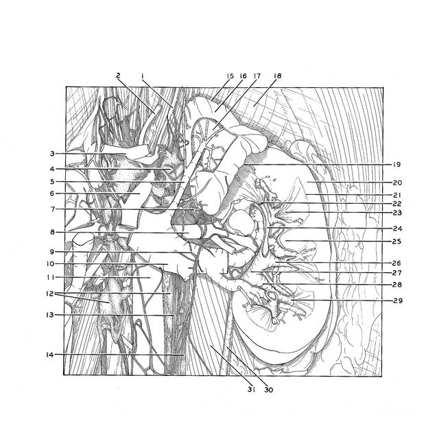
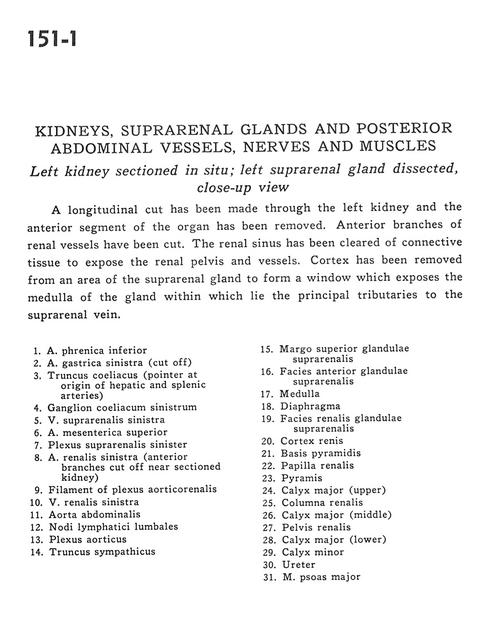
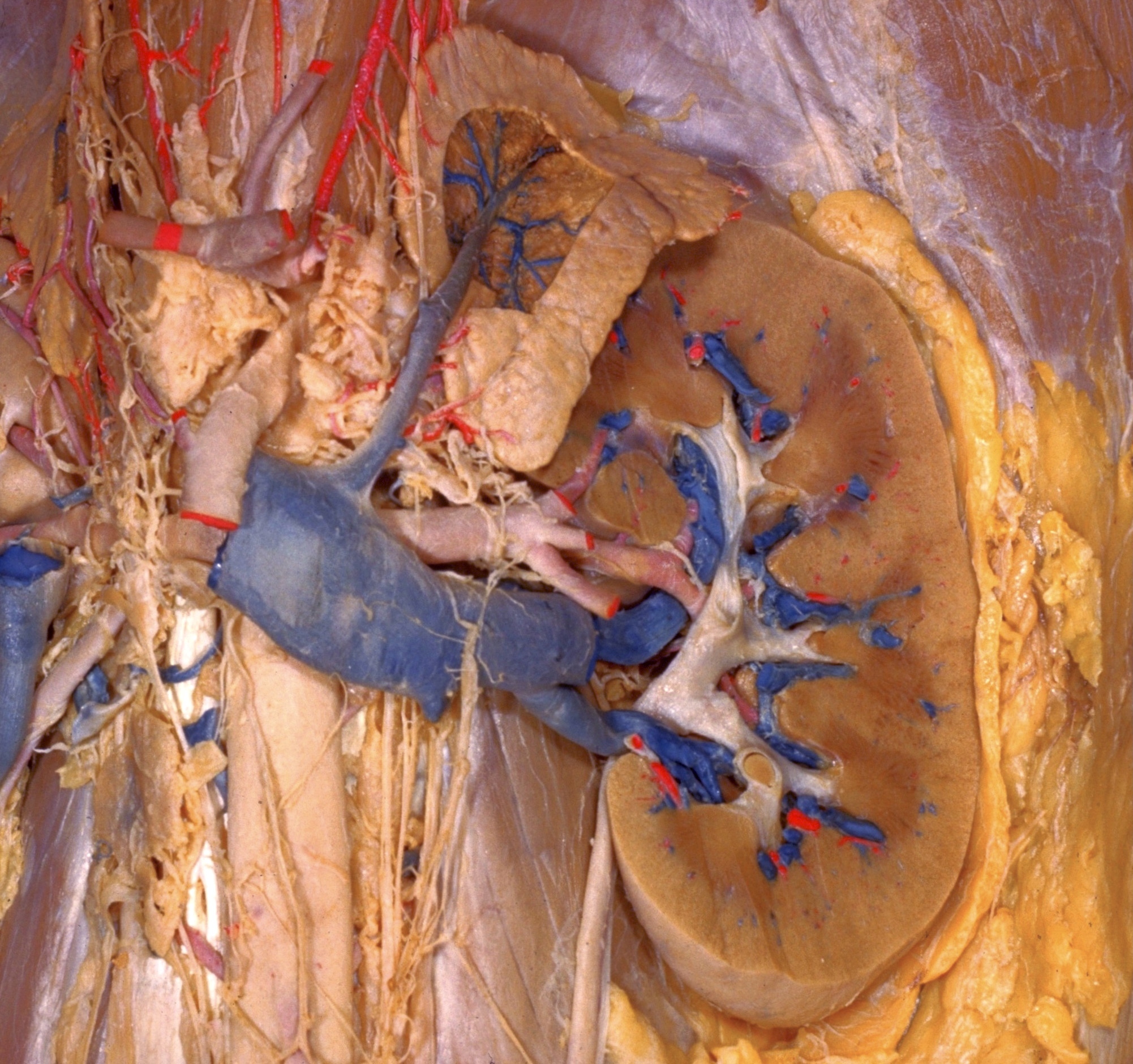
Kidneys, suprarenal glands and posterior abdominal vessels, nerves and muscles
Left kidney sectioned in situ; left suprarenal gland dissected, close-up view
A longitudinal cut has been made through the left kidney and the anterior segment of the organ has been removed. Anterior branches of renal vessels have been cut. The renal sinus has been cleared of connective tissue to expose the renal pelvis and vessels. Cortex has been removed from an area of the suprarenal gland to form a window which exposes the medulla of the gland within which lie the principal tributaries to the suprarenal vein.
- Inferior phrenic artery
- Left gastric artery (cut off)
- Celiac trunk (pointer at origin of hepatic and splenic arteries)
- Left celiac ganglion
- Left suprarenal vein
- Superior mesenteric artery
- Left suprarenal plexus
- Left renal artery (anterior branches cut off near sectioned kidney)
- Filament of aorticorenal plexus
- Left renal vein
- Abdominal aorta
- Lumbar lymph nodes
- Aortic plexus
- Sympathetic trunk
- Superior margin of suprarenal gland
- Anterior surface of suprarenal gland
- Medulla
- Diaphragm
- Renal surface of suprarenal gland
- Cortex of kidney
- Base of pyramid
- Renal papilla
- Pyramid
- Calyx major (upper)
- Renal column
- Calyx major (middle)
- Renal pelvis
- Calyx major (lower)
- Minor calyx
- Ureter
- Psoas major muscle