Kidneys, suprarenal, glands and posterior abdominal vessels, nerves and muscles
Fascial relations of left kidney, close--up anterior view
Stanford holds the copyright to the David L. Bassett anatomical images and has assigned
Creative Commons license Attribution-Share
Alike 4.0 International to all of the images.
For additional information regarding use and permissions,
please contact Dr. Drew Bourn at dbourn@stanford.edu.
Image #149-1
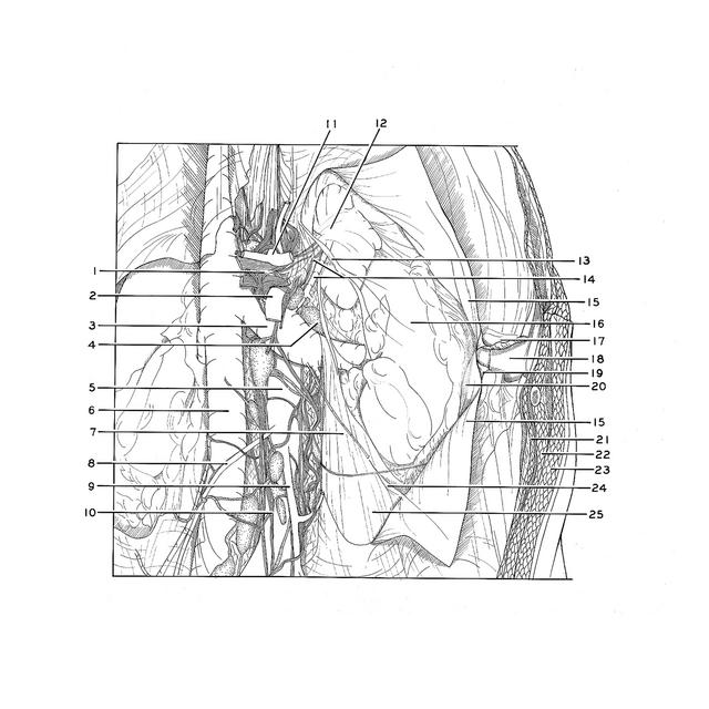
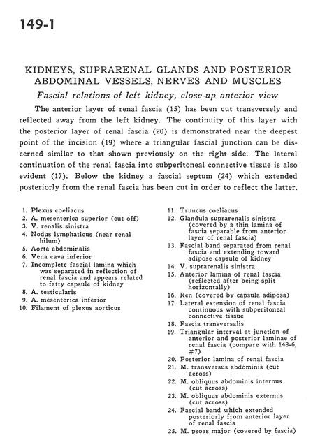
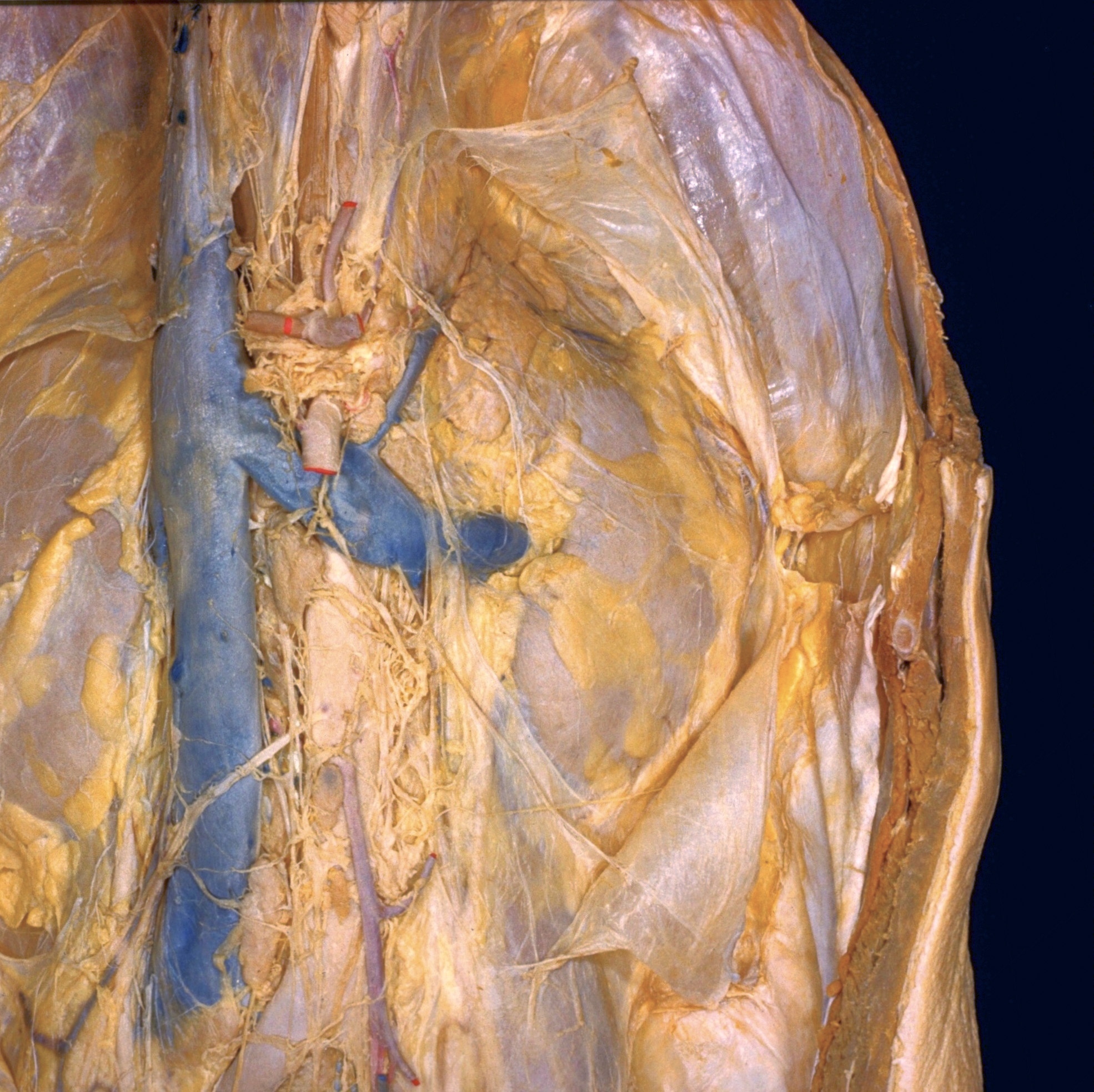
Kidneys, suprarenal, glands and posterior abdominal vessels, nerves and muscles
Fascial relations of left kidney, close--up anterior view
The anterior part of renal fascia (15) has been cut transversely and reflected away from the left kidney. The continuity of this layer with the posterior layer of renal fascia (20) id demonstrated near the deepest point of incision (19) where a triangular fascial junction can be discerned similar to that shown previously on the right side. The lateral continuation of the renal fascia into subperitoneal connective tissue is also evident (17). Below the kidney a fascial septum (24) which extended posteriorly from the renal fascia has been cut in order to reflect the latter.
- Celiac plexus
- Superior mesenteric artery (cut off)
- Left renal vein
- Lymph node (near renal hilum)
- Abdominal aorta
- Inferior vena cava
- Incomplete fascial lamina which was separated in reflection of renal fascia and appears related to fatty capsule of kidney
- Testicular artery
- Inferior mesenteric artery
- Filament of aortic plexus
- Celiac trunk
- Left suprarenal gland (covered by a thin lamina of fascia separable from anterior layer of renal fascia)
- Fascial band separated from renal fascia and extending toward adipose capsule of kidney
- Left suprarenal vein
- Anterior layer of renal fascia (reflected after being split horizontally)
- Kidney (covered by fatty capsule)
- Lateral extension of renal fascia continuous with subperitoneal connective tissue
- Transversalis fascia
- Triangular interval at junction of anterior and posterior laminae of renal fascia (compare with 148-6#7)
- Posterior lamina of renal fascia
- Transversus abdominis muscle (cut across)
- Internal oblique muscle (cut across)
- External oblique muscle (cut across)
- Fascial band which extended posteriorly from anterior layer of renal fascia
- Psoas major muscle (covered by fascia)