Exploration of liver, gall bladder, pancreas, duodenum and spleen
General view of organs in situ, stomach elevated
Stanford holds the copyright to the David L. Bassett anatomical images and has assigned
Creative Commons license Attribution-Share Alike 4.0 International to all of the images.
For additional information regarding use and permissions,
please contact Dr. Drew Bourn at dbourn@stanford.edu.
Image #145-4
 |  |
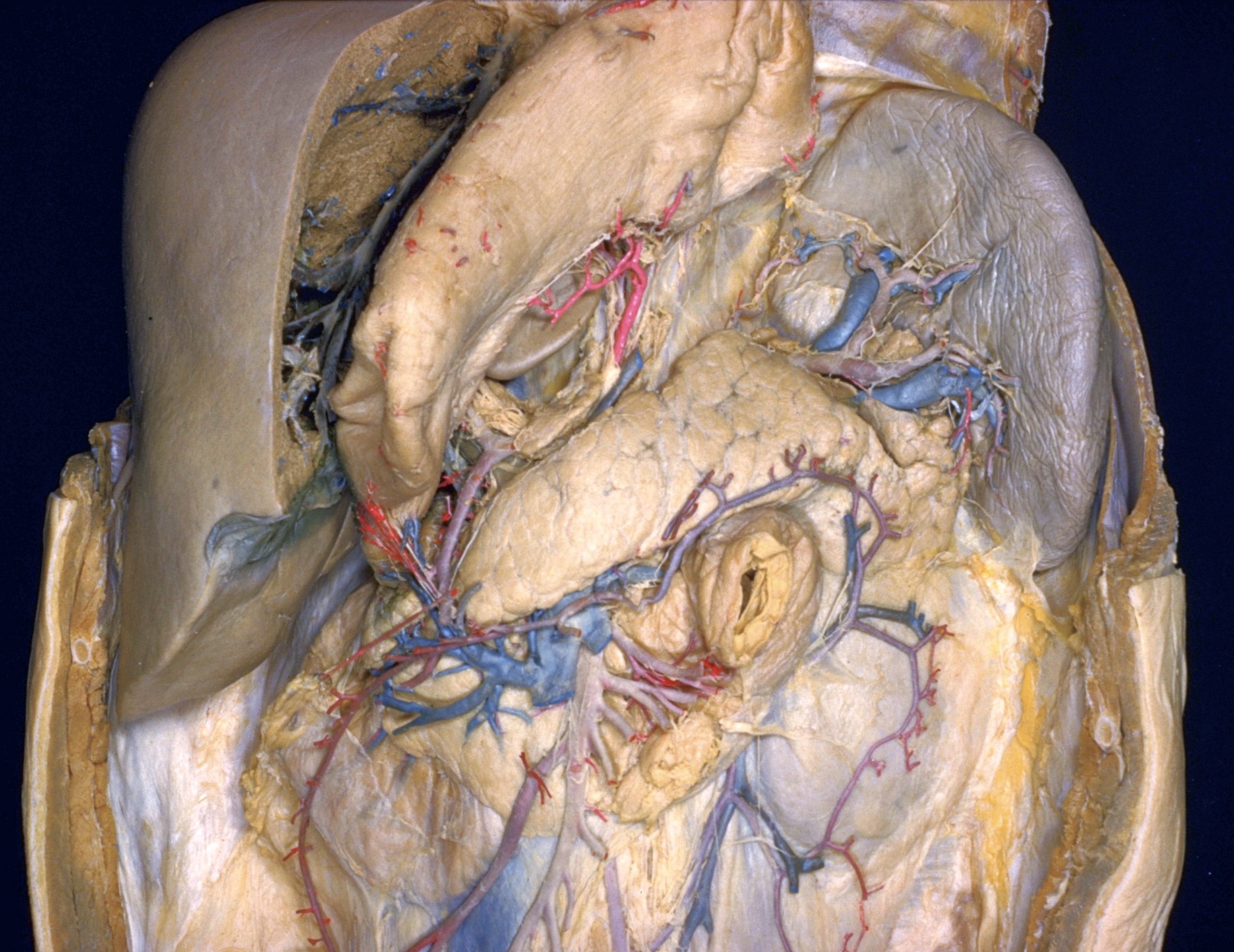
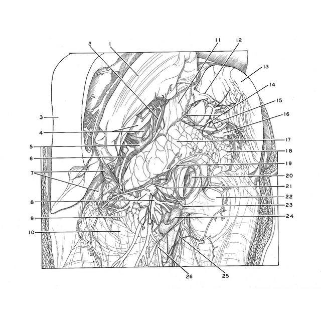
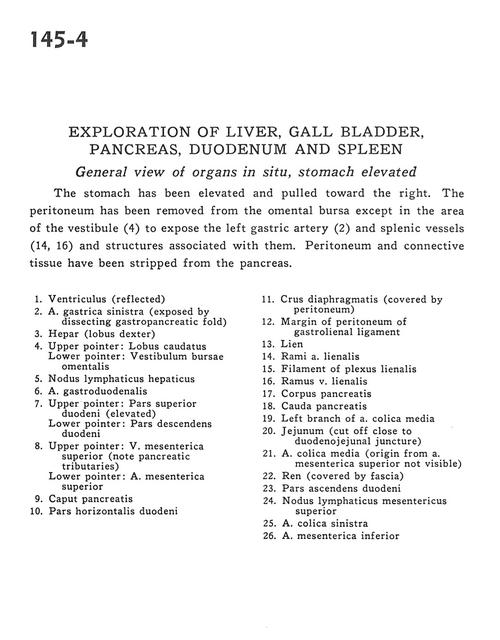
 | | Exploration of liver, gall bladder, pancreas, duodenum and spleen | | General view of organs in situ, stomach elevated | | The stomach has been elevated and pulled toward the right. The peritoneum has been removed from the omental bursa except in the area of the vestibule (4) to expose the left gastric artery (2) and splenic vessels (14,16) and structures associated with them. Peritoneum and connective tissue have been stripped from the pancreas. | | 1
.
| Stomach (reflected) | | 2
.
| Left gastric artery (exposed by dissecting gastropancreatic fold) | | 3
.
| Liver (right lobe) | | 4
.
| Upper pointer: Caudate lobe Lower pointer: Vestibule of omental bursa | | 5
.
| Hepatic lymph node | | 6
.
| Gastroduodenal artery | | 7
.
| Upper pointer: Superior part of duodenum (elevated) Lower pointer: Descending part of duodenum | | 8
.
| Upper pointer: Superior mesenteric vein (note pancreatic tributaries) Lower pointer: Superior mesenteric artery | | 9
.
| Head of pancreas | | 10
.
| Horizontal part of duodenum | | 11
.
| Crus of diaphragm (covered by peritoneum) | | 12
.
| Margin of peritoneum of gastrolienal ligament | | 13
.
| Spleen | | 14
.
| Branches of splenic artery | | 15
.
| Filament of splenic plexus | | 16
.
| Branch of splenic vein | | 17
.
| Body of pancreas | | 18
.
| Tail of pancreas | | 19
.
| Left branch of middle colic artery | | 20
.
| Jejunum (cut off close to duodenojejunal juncture) | | 21
.
| Middle colic artery (origin from superior mesenteric artery not visible) | | 22
.
| Kidney (covered by fascia) | | 23
.
| Ascending part of duodenum | | 24
.
| Superior mesenteric lymph node | | 25
.
| Left colic artery | | 26
.
| Inferior mesenteric artery |
|
|


