Exploration of peritoneal cavity
Upper abdominal organs in situ
Stanford holds the copyright to the David L. Bassett anatomical images and has assigned
Creative Commons license Attribution-Share
Alike 4.0 International to all of the images.
For additional information regarding use and permissions,
please contact Dr. Drew Bourn at dbourn@stanford.edu.
Image #138-6
Exploration of peritoneal cavity
Upper abdominal organs in situ
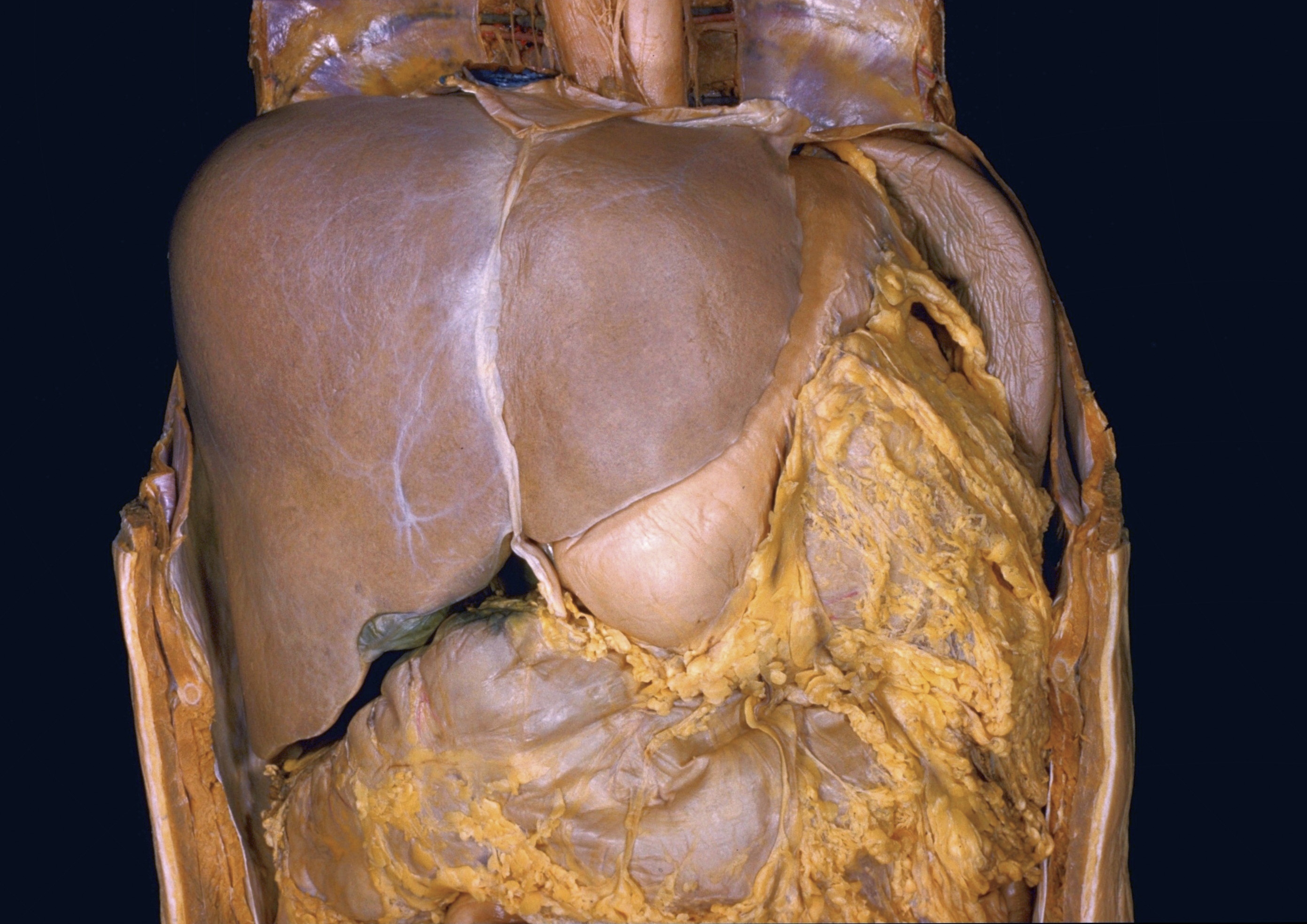
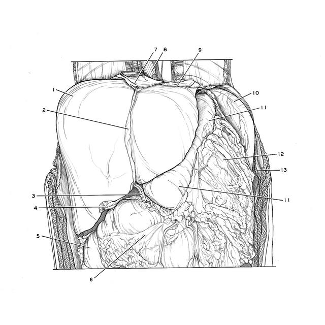
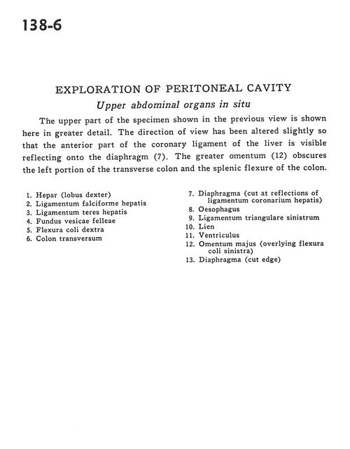
The upper part of the specimen shown in the previous view is shown here in great detail. The direction of view has been altered slightly so that the anterior part of the coronary ligament of the liver is visible reflecting onto the diaphragm(7). The greater omentum (12) obscures the left portion of the transverse colon and the splenic flexure of the colon.
- Liver (right lobe)
- Falciform ligament of liver
- Ligamentum teres (of liver)
- Fundus gallbladder
- Right colic flexure
- Transverse colon
- Diaphragm (cut at reflections of coronary ligament of liver)
- Esophagus
- Left triangular ligament
- Spleen
- Stomach
- Greater omentum (overlying left colic flexure)
- Diaphragm (cut edge)