Dissection of male inguinal region and spermatic cord
Internal oblique and cremaster muscles
Stanford holds the copyright to the David L. Bassett anatomical images and has assigned
Creative Commons license Attribution-Share
Alike 4.0 International to all of the images.
For additional information regarding use and permissions,
please contact Dr. Drew Bourn at dbourn@stanford.edu.
Image #136-1
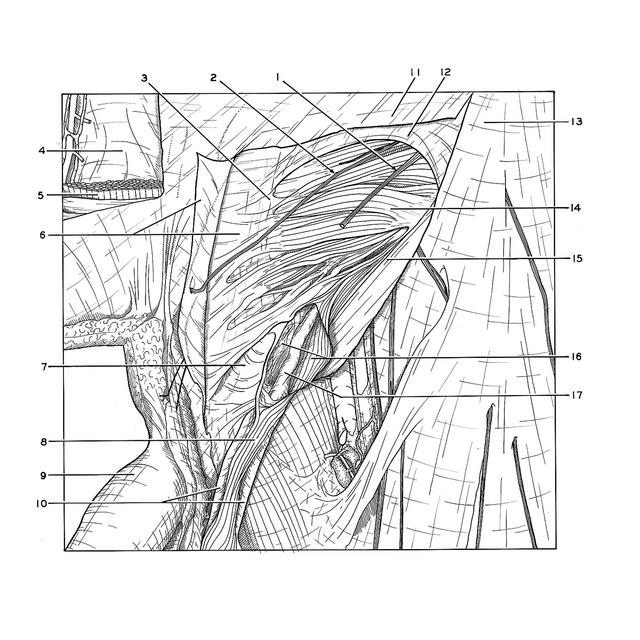
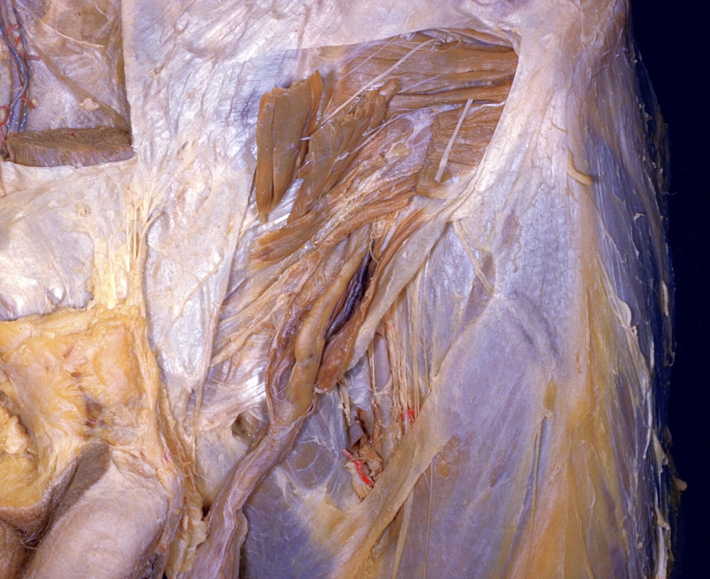
Dissection of male inguinal region and spermatic cord
Internal oblique and cremaster muscles
The aponeurosis of the external oblique (2) has been excised above and medial to the inguinal canal. Fascia has been cut away from the underlying internal oblique muscle. The origin of the cremaster muscle (16) from the lower border of the internal oblique is visible. The cremaster has been split to demonstrate the manner in which it encases the inner structures of the spermatic cord (17) in their passage along the inguinal canal.
- Ilioinguinal nerve
- Iliohypogastric nerve
- Left rectus abdominis muscle (pointer indicates lateral margin of muscle visible through rectus sheath)
- Transversalis fascia
- Right rectus abdominis muscle (cut across)
- Sheath of rectus abdominis muscle (upper pointer indicates aponeurosis of external oblique, split and reflected from sheath)
- Reflected inguinal ligament (exposed by lateral displacement of spermatic cord)
- Spermatic cord
- Superficial fascia of the penis (dartos)
- Cremasteric fascia (reflected)
- Aponeurosis external oblique muscle
- Fascia of internal oblique muscle
- Location of anterior superior iliac spine
- Internal oblique muscle
- Inguinal ligament
- Cremaster muscle (split open)
- Internal spermatic fascia (enclosing ductus deferens and associated structures)