X CLOSE

Exploration of the brain from its lateral aspect

General lateral view of brain

Stanford holds the copyright to the David L. Bassett anatomical images and has assigned Creative Commons license Attribution-Share Alike 4.0 International to all of the images.
For additional information regarding use and permissions, please contact Dr. Drew Bourn at dbourn@stanford.edu.

Image #13-1
Bassett ImageDiagram Image
Legend Image
Exploration of the brain from its lateral aspect
General lateral view of brain
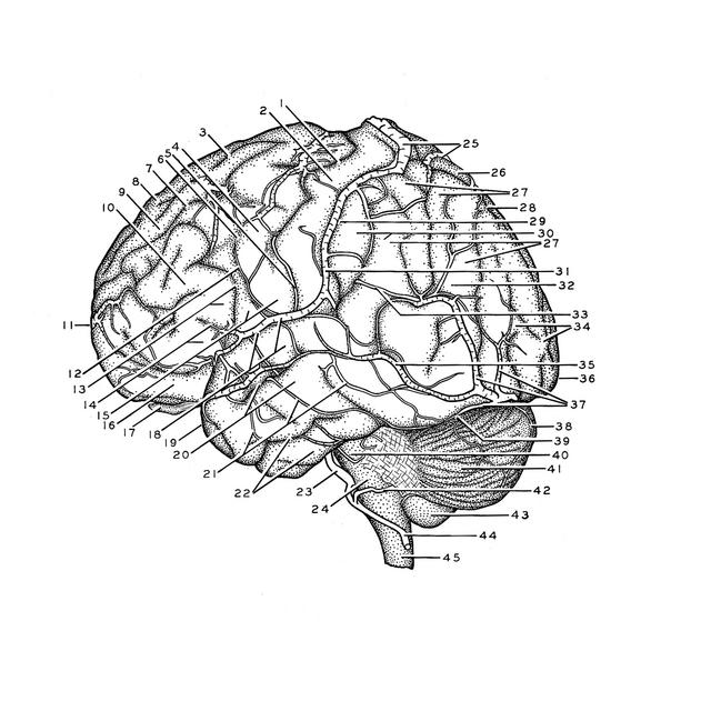
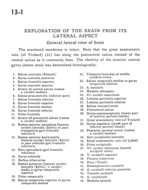
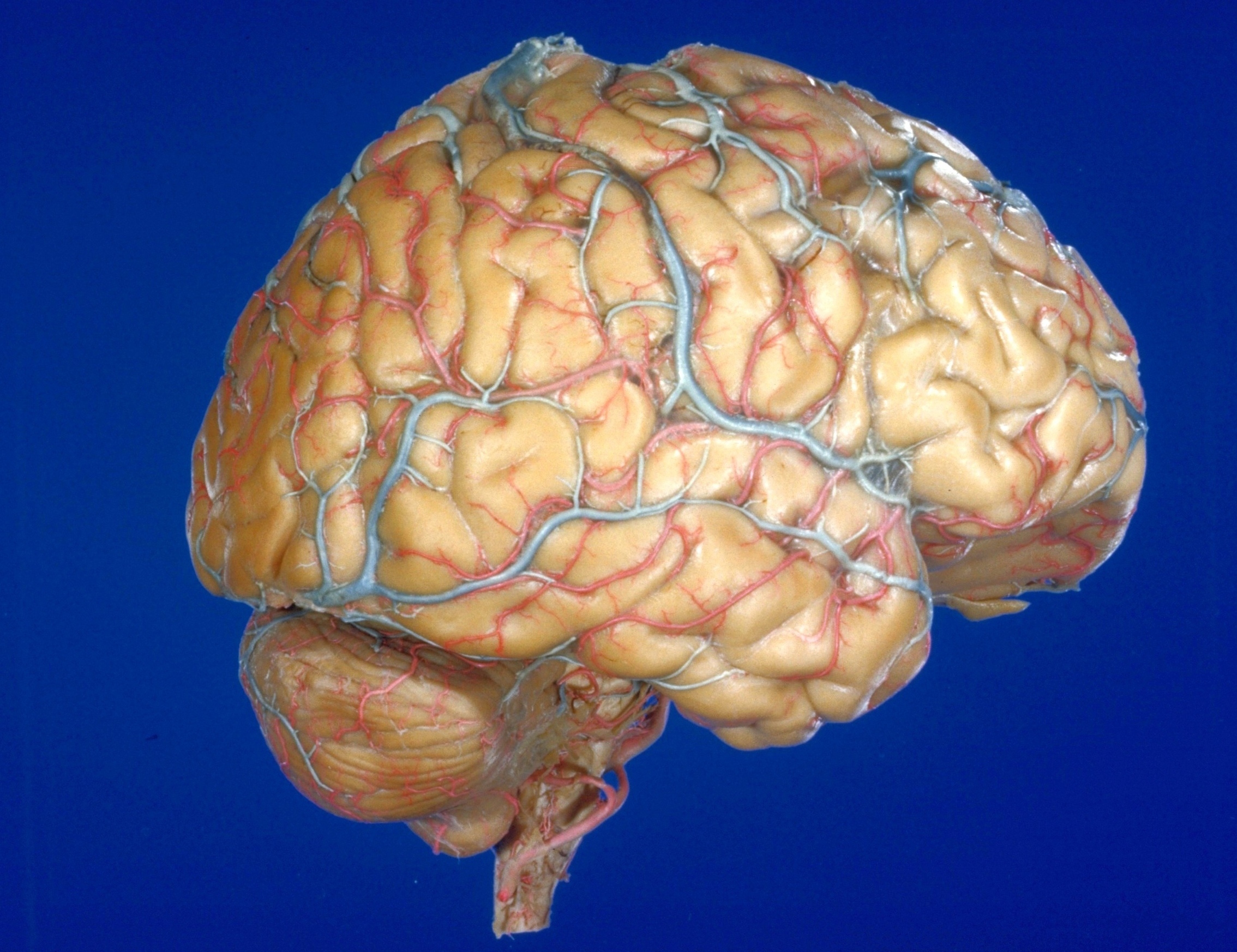
The arachnoid membrane is intact. Note that the great anastomotic vein (of Trolard) (31) lies along the postcentral sulcus instead of the central sulcus as it commonly does. The identity of the anterior central gyrus (motor area) was determined histologically.
1 . Central sulcus (of Rolando)
2 . Postcentral gyrus
3 . Superior frontal sulcus
4 . Precentral gyrus
5 . Artery of central sulcus (branch middle cerebral artery)
6 . Precentral sulcus (inferior part)
7 . Inferior frontal sulcus
8 . Superior frontal gyrus
9 . Medial frontal gyrus
10 . Inferior frontal gyrus
11 . Frontal pole
12 . Artery of precentral sulcus (branch middle cerebral artery)
13 . Anterior ascending branch of lateral cerebral fissure and triangular part of inferior frontal gyrus
14 . Anterior horizontal branch lateral cerebral fissure and orbital part inferior frontal gyri
15 . Operculum of inferior frontal gyrus
16 . Orbital gyrus
17 . Olfactory bulb
18 . Posterior branch of lateral cerebral fissure, middle cerebral vein and superior temporal gyrus
19 . Temporal pole
20 . Superior temporal sulcus and medial temporal gyrus
21 . Temporal branches of middle cerebral artery
22 . Middle temporal sulcus and inferior temporal gyrus
23 . Basilar artery
24 . Medulla oblongata
25 . Superior cerebral veins
26 . Superior parietal lobule
27 . Inferior parietal lobule
28 . Interparietal sulcus
29 . Postcentral sulcus
30 . Supramarginal gyrus (lower part of inferior parietal lobule)
31 . Great anastomotic vein (of Trolard)
32 . Angular gyrus (lower part of inferior parietal lobule)
33 . Posterior parietal artery (branch middle cerebral artery)
34 . Lateral occipital gyri
35 . Small anastomotic vein (of Labbé)
36 . Occipital pole
37 . Inferior cerebral veins (lateral occipital veins)
38 . Superior cerebellar vein
39 . Transverse fissure
40 . Pons
41 . Cerebellar hemisphere
42 . Posterior inferior cerebellar artery
43 . Cerebellar tonsil
44 . Vertebral artery
45 . Spinal medulla