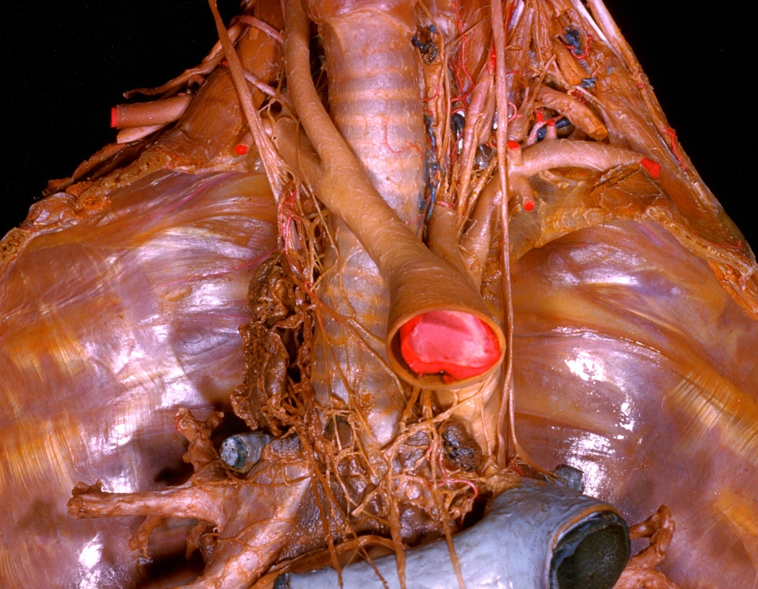
Dissection of mediastinum and paravertebral structures
Superior mediastinum.
Stanford holds the copyright to the David L. Bassett anatomical images and has assigned
Creative Commons license Attribution-Share
Alike 4.0 International to all of the images.
For additional information regarding use and permissions,
please contact Dr. Drew Bourn at dbourn@stanford.edu.
Image #127-5
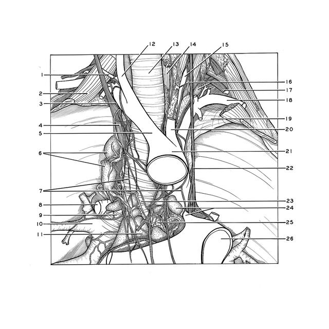
Dissection of mediastinum and paravertebral structures
Superior mediastinum.
The aortic arch has been elevated and the pulmonary arteries have been retracted downward.
- Transverse scapular artery
- Anterior scalene muscle
- Recurrent laryngeal nerve
- Right vagus nerve
- Brachiocephalic trunk
- Superior tracheobronchial lymph nodes
- Cardiac plexus
- Azygos vein (cut off)
- Right main bronchus (note filaments of pulmonary plexus along bronchus)
- Bronchus of upper right lobe
- Inferior tracheobronchial lymph node
- Common carotid artery
- Trachea
- Esophagus
- Vertebral artery
- Stellate ganglion
- Cervical nerve VIII
- Subclavian artery
- Cupula pleurae
- Left common carotid artery
- Aortic arch (elevated)
- Vagus nerve left
- Tracheal bifurcation
- Upper pointer: Superior tracheobronchial lymph nodes Lower pointer: Ligamentum arteriosum
- Left main bronchus
- Pulmonary trunk (retracted downward)