Dissection of lungs in situ
Right lung.
Stanford holds the copyright to the David L. Bassett anatomical images and has assigned
Creative Commons license Attribution-Share
Alike 4.0 International to all of the images.
For additional information regarding use and permissions,
please contact Dr. Drew Bourn at dbourn@stanford.edu.
Image #124-4
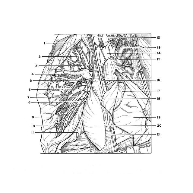

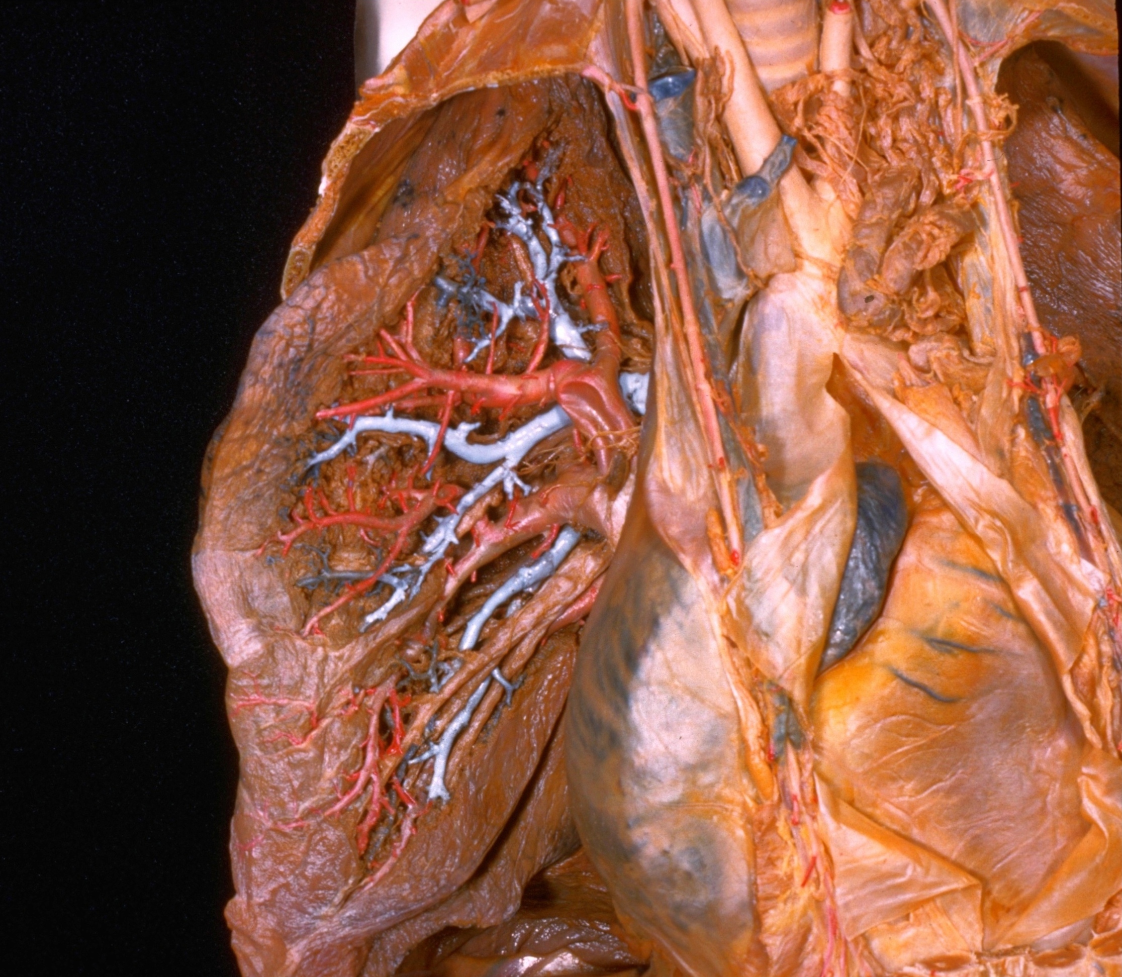
Dissection of lungs in situ
Right lung.
The dissection has been extended from that shown in the previous view to include the anterior and apical segments of the upper lobe.
- Upper lobe right lung
- Apical segmental bronchus (pointer on apical branch)
- Apical branch right pulmonary artery
- Superior intersegmental vein
- Upper pointer: Anterior branch right pulmonary artery Lower pointer Intersegmental vein
- Anterior segmental bronchus (pointer on anterior branch)
- Branch to middle lobe right pulmonary artery
- Medial segmental bronchus of middle lobe
- Division between upper and middle lobes (no horizontal fissure on medial aspect of specimen)
- Middle lobe right lung
- Inferior lobe right lung
- Brachiocephalic trunk and right brachiocephalic vein
- Anterior mediastinal lymph node and lymph vessel
- Left brachiocephalic vein (cut off)
- Aortic arch
- Internal thoracic artery and vein and lymph vessel
- Right auricle
- Pericardium (reflected)
- Right ventricle
- Right atrium (covered by pleura and pericardium)
- Transversus thoracis muscle (cut off)