Dissection of pericardium and heart in situ
Heart exposed within pericardial activity
Stanford holds the copyright to the David L. Bassett anatomical images and has assigned
Creative Commons license Attribution-Share
Alike 4.0 International to all of the images.
For additional information regarding use and permissions,
please contact Dr. Drew Bourn at dbourn@stanford.edu.
Image #116-7
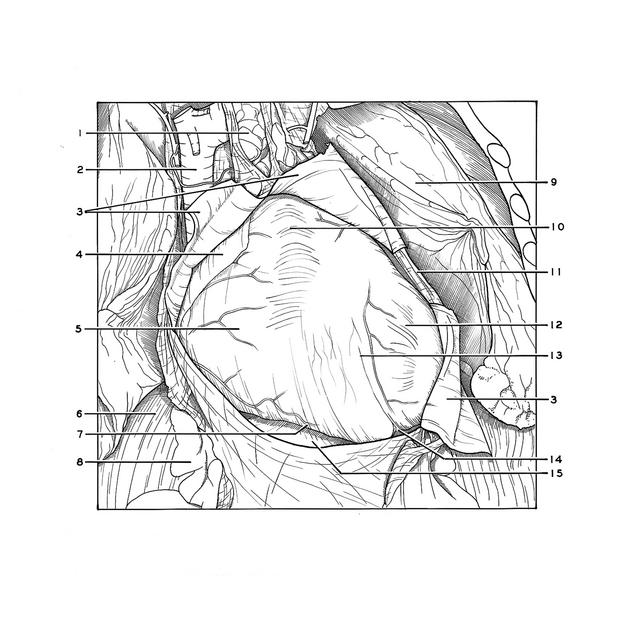
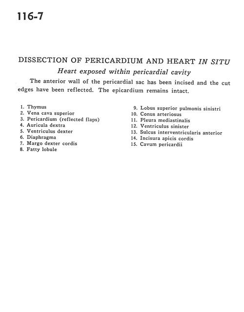
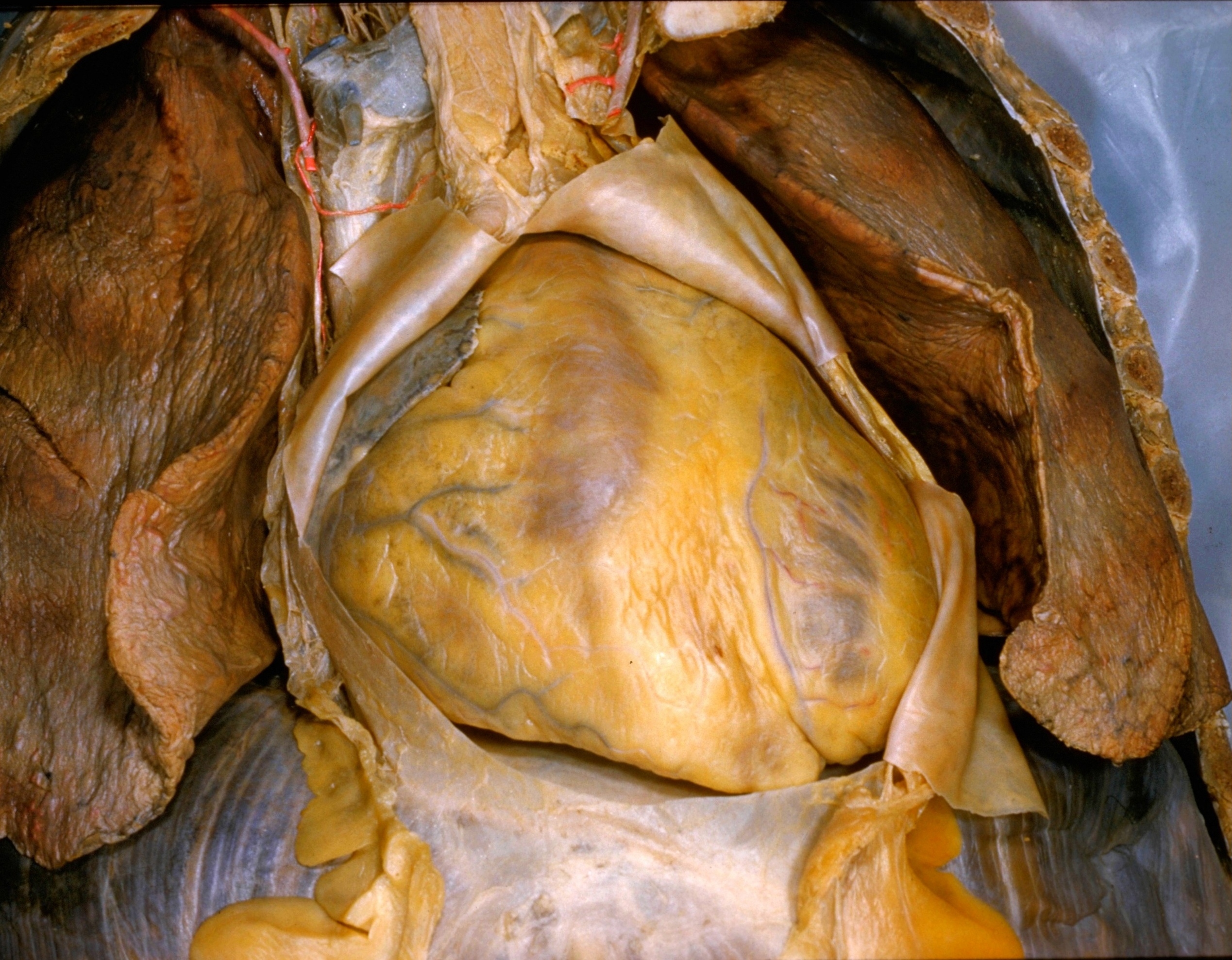
Dissection of pericardium and heart in situ
Heart exposed within pericardial activity
The anterior wall of the pericardial sac has been incised and the cut edges have been reflected. The epicardium remains intact.
- Thymus
- Superior vena cava
- Pericardium (reflected flaps)
- Right auricle
- Right ventricle
- Diaphragm
- Right margin of heart
- Fatty lobule
- Upper lobe left lung
- Conus arteriosus
- Mediastinal pleura
- Left ventricle
- Anterior interventricular sulcus
- Arcuate margin of heart
- Pericardial cavity