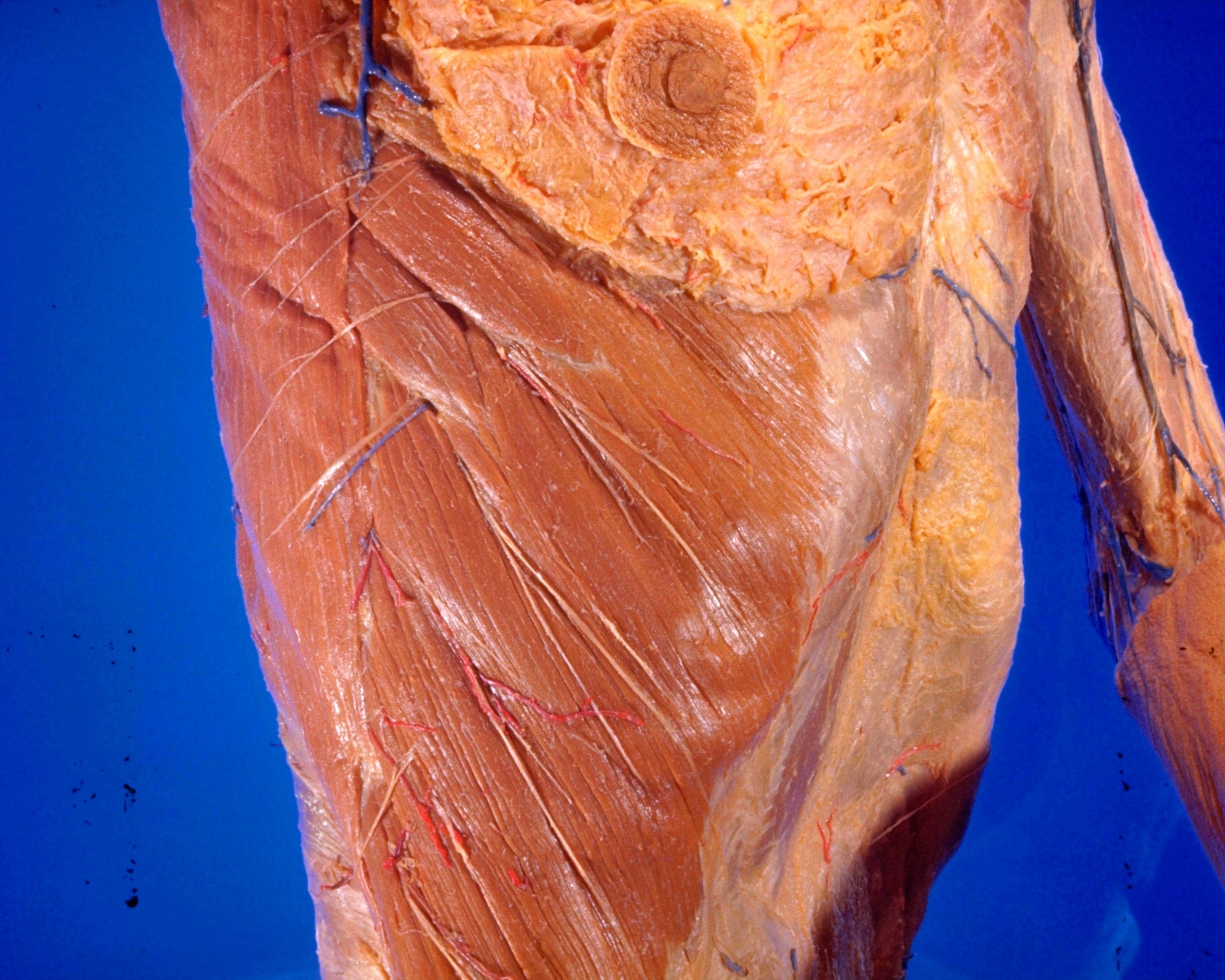
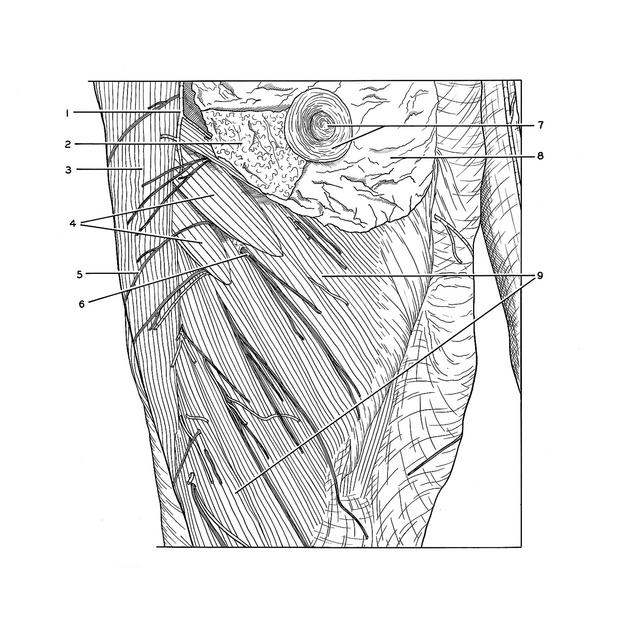
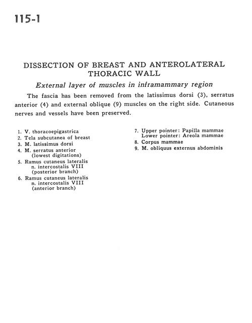
Dissection of breast and anterolateral thoracic wall
External layer of muscles in inframammary region
Stanford holds the copyright to the David L. Bassett anatomical images and has assigned
Creative Commons license Attribution-Share
Alike 4.0 International to all of the images.
For additional information regarding use and permissions,
please contact Dr. Drew Bourn at dbourn@stanford.edu.
Image #115-1
Dissection of breast and anterolateral thoracic wall
External layer of muscles in inframammary region
The fascia has been removed from the latissimus dorsi (3), serratus anterior (4) and external oblique (9) muscles on the right side. Cutaneous nerves and vessels have been preserved.
- Thoracoepigastric vein
- Superficial fascia of breast
- Latissimus dorsi muscle
- Serratus anterior muscle (lowest digitations)
- Lateral cutaneous branch intercostal nerve VIII (posterior branch)
- Lateral cutaneous branch intercostal nerve VIII (anterior branch)
- Upper pointer: Nipple (mammary papilla) Lower pointer: Areola
- Mammary body
- External abdominal oblique muscle