Dissection of breast and anterolateral thoracic wall
Right breast dissected in situ
Stanford holds the copyright to the David L. Bassett anatomical images and has assigned
Creative Commons license Attribution-Share
Alike 4.0 International to all of the images.
For additional information regarding use and permissions,
please contact Dr. Drew Bourn at dbourn@stanford.edu.
Image #114-6
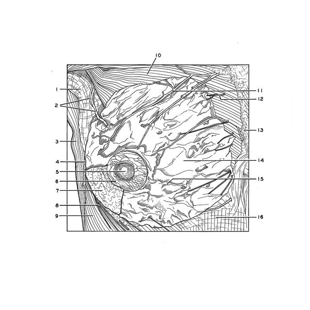
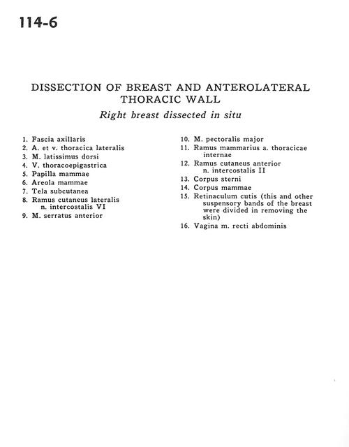
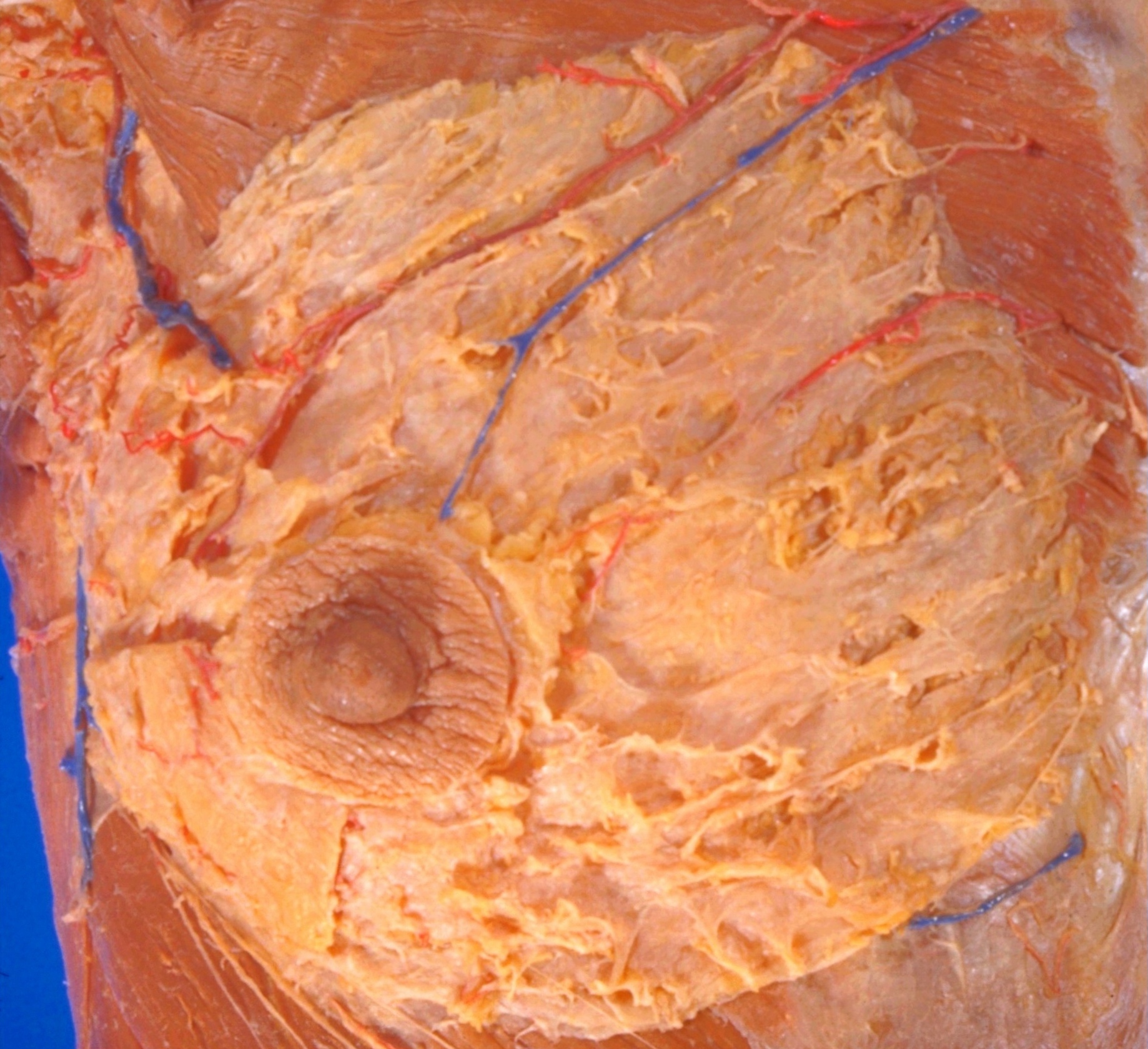
Dissection of breast and anterolateral thoracic wall
Right breast dissected in situ
- Axillary fascia
- Lateral thoracic artery and vein
- Latissimus dorsi muscle
- Thoracoepigastric vein
- Nipple (mammary papilla)
- Areola
- Superficial fascia
- Lateral cutaneous branch intercostal nerve VI
- Serratus anterior muscle
- Pectoralis major muscle
- Mammary branch internal thoracic artery
- Anterior cutaneous branch intercostal nerve II
- Body of sternum
- Mammary body
- Cutaneous ligaments (this and other suspensory bands of the breast were divided in removing the skin)
- Sheath of rectus abdominis muscle