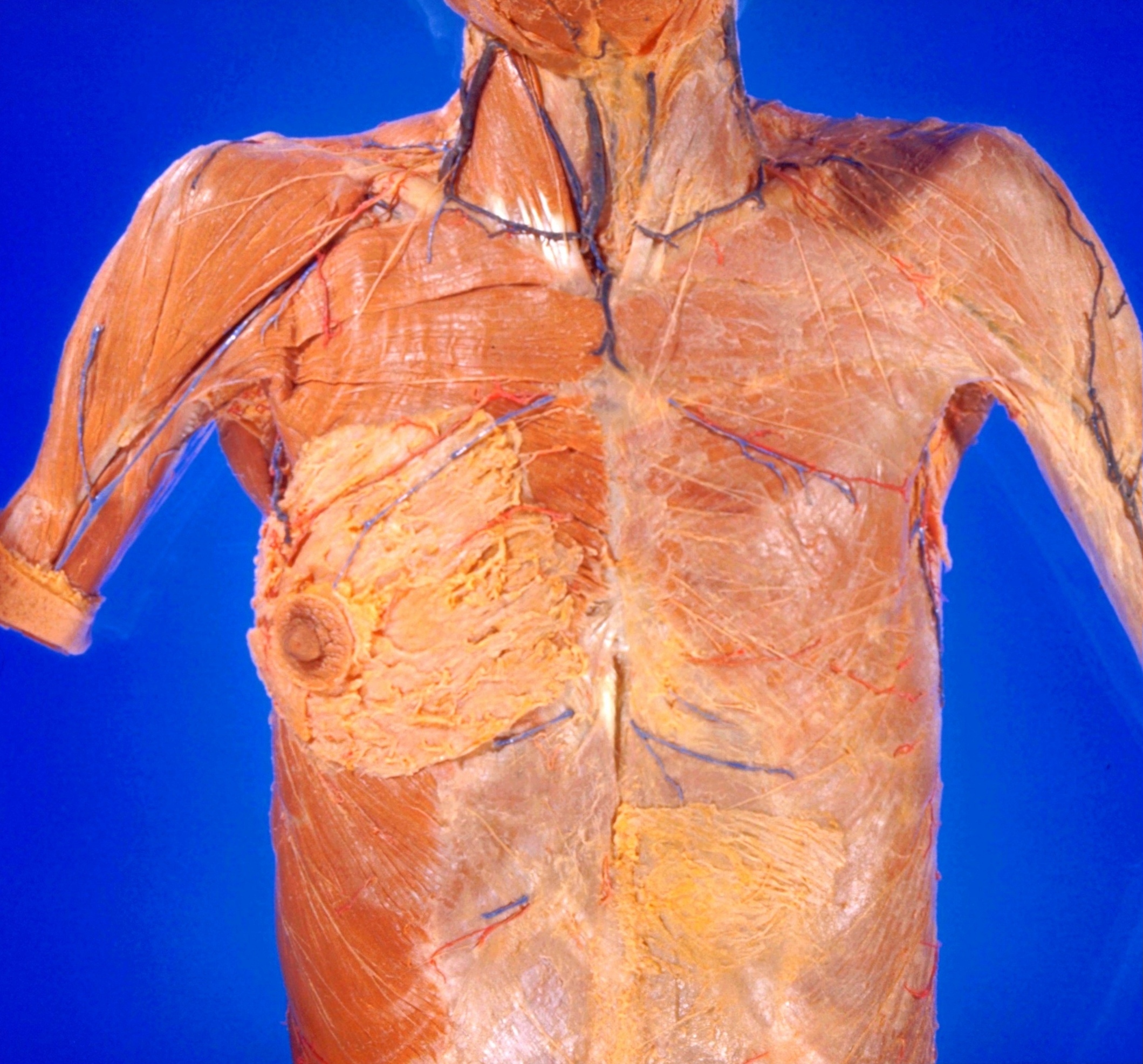
Dissection of breast and anterolateral thoracic wall
Superficial vessels and nerves; breast; pectoral fascia and muscles
Stanford holds the copyright to the David L. Bassett anatomical images and has assigned
Creative Commons license Attribution-Share
Alike 4.0 International to all of the images.
For additional information regarding use and permissions,
please contact Dr. Drew Bourn at dbourn@stanford.edu.
Image #114-5
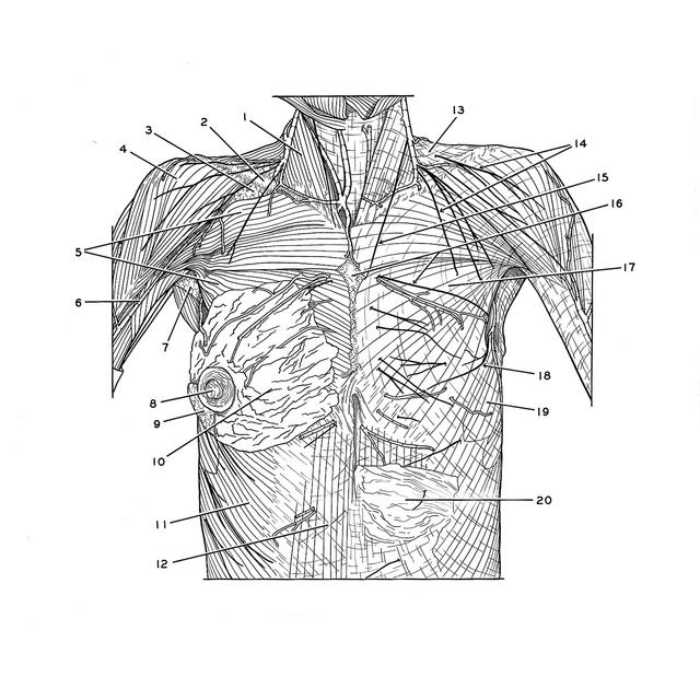
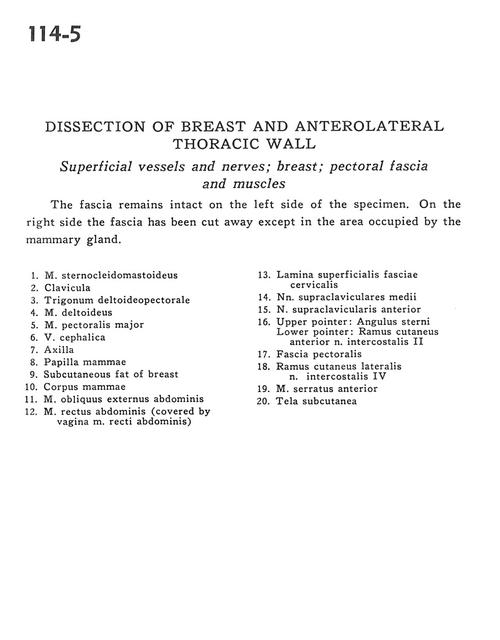
Dissection of breast and anterolateral thoracic wall
Superficial vessels and nerves; breast; pectoral fascia and muscles
The fascia remains intact on the left side of the specimen. On the right side the fascia has been cut away except in the area occupied by the mammary gland.
- Sternocleidomastoid muscle
- Clavicle
- Deltopectoral triangle
- Deltoid muscle
- Pectoralis major muscle
- Cephalic vein
- Axilla
- Nipple (mammary papilla)
- Subcutaneous fat of breast
- Mammary body
- External abdominal oblique muscle
- Rectus abdominis muscle (covered by sheath of rectus abdominis muscle)
- Superficial cervical fascia
- Middle supraclavicular nerves
- Anterior supraclavicular nerve
- Upper pointer: Sternal angle Lower pointer: Anterior cutaneous branch intercostal nerve II
- Pectoral fascia
- Lateral cutaneous branch intercostal nerve IV
- Serratus anterior muscle
- Superficial fascia