Joints of right index finger
Capsule and ligaments of metacarpophalangeal joint, medial view
Stanford holds the copyright to the David L. Bassett anatomical images and has assigned
Creative Commons license Attribution-Share
Alike 4.0 International to all of the images.
For additional information regarding use and permissions,
please contact Dr. Drew Bourn at dbourn@stanford.edu.
Image #111-2
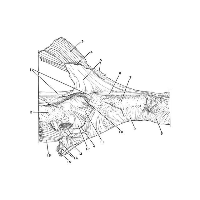
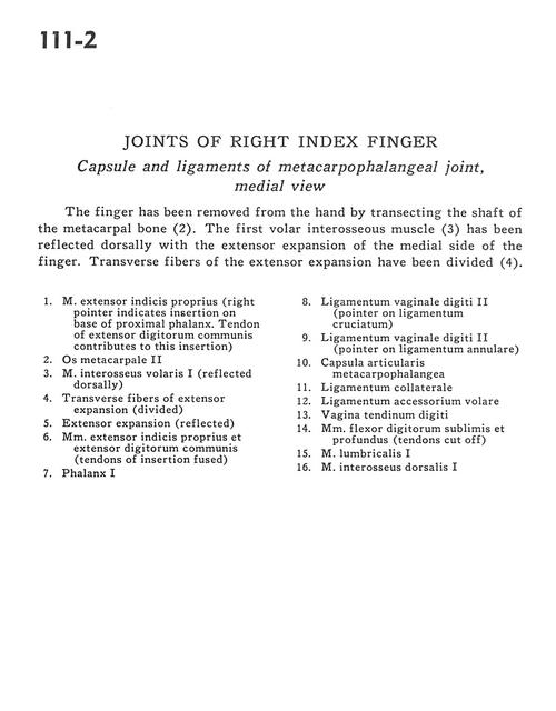
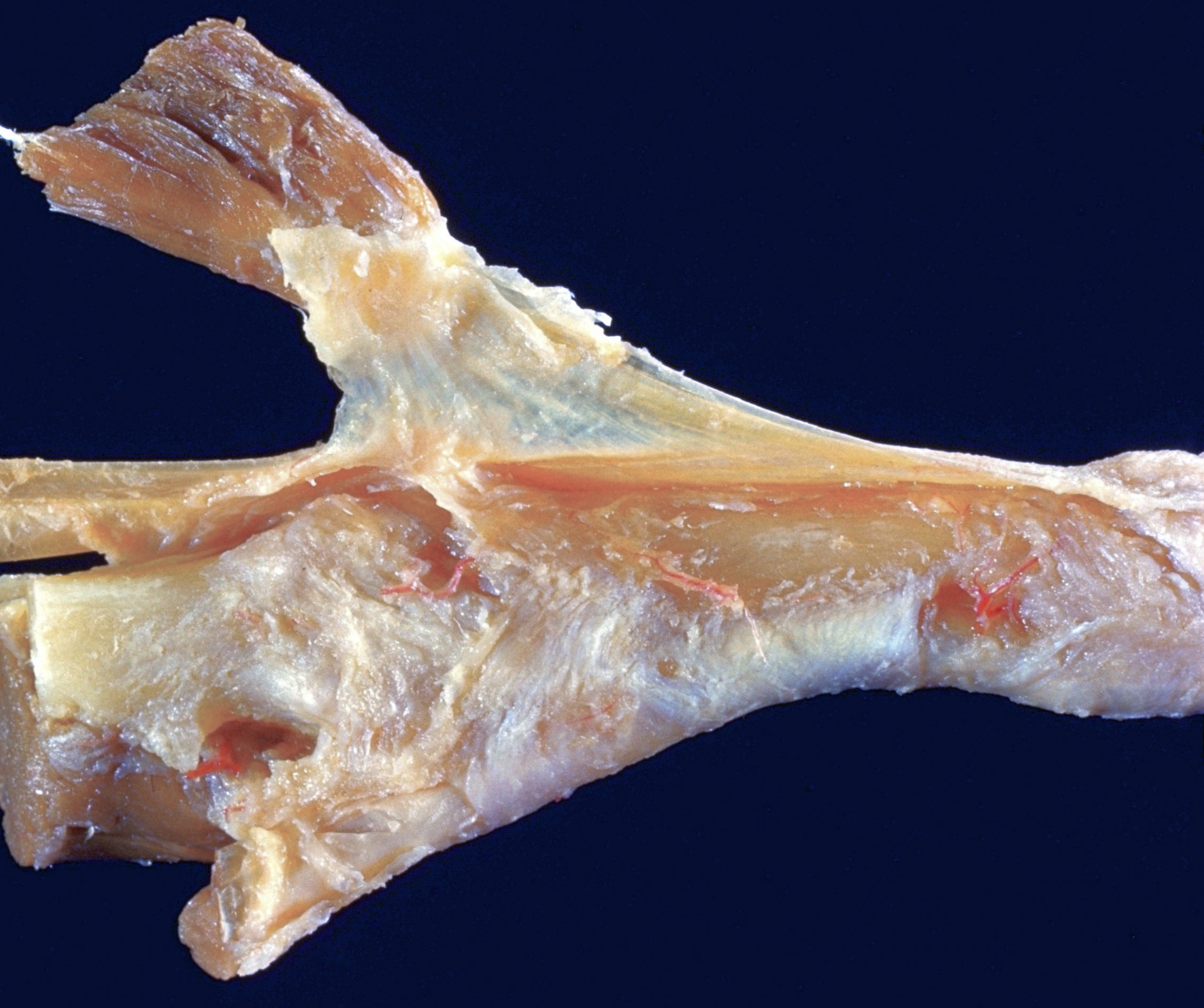
Joints of right index finger
Capsule and ligaments of metacarpophalangeal joint, medial view
The finger has been removed from the hand by transecting the shaft of the metacarpal bone (2). The first volar interosseous muscle (3) has been reflected dorsally with the extensor expansion of the medial side of the finger. Transverse fibers of the extensor expansion have been divided (4).
- Extensor indicis muscle (right pointer indicates insertion on base of proximal phalanx. Tendon of extensor digitorum contributes to this insertion)
- Metacarpal II
- Anterior interosseous muscle I (reflected dorsally)
- Transverse fibers of extensor expansion (divided)
- Extensor expansion (reflected)
- Extensor indicis muscle and common extensor digitorum muscle (tendons of insertion fused)
- Phalanx I
- Ligament of digital sheath II (pointer on cruciate ligament)
- Ligament of digital sheath II (pointer on annular ligament)
- Metacarpophalangeal joint capsule
- Collateral ligament
- Anterior accessory ligament
- Tendon of digital sheath
- Flexor digitorum superficialis and profundus muscles (tendons cut off)
- Lumbrical muscle I
- Dorsal interosseous muscle I