Exploration of the brain from its superior aspect
Internal capsule exposed by removing head and body of caudate nucleus
Stanford holds the copyright to the David L. Bassett anatomical images and has assigned
Creative Commons license Attribution-Share
Alike 4.0 International to all of the images.
For additional information regarding use and permissions,
please contact Dr. Drew Bourn at dbourn@stanford.edu.
Image #11-5
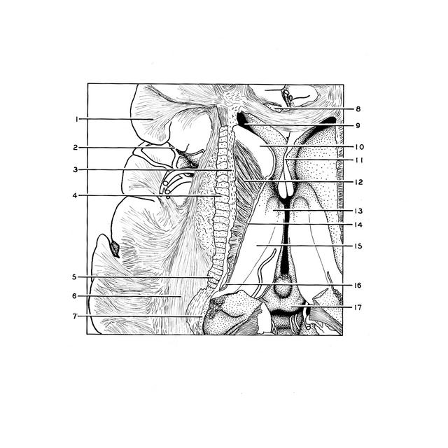
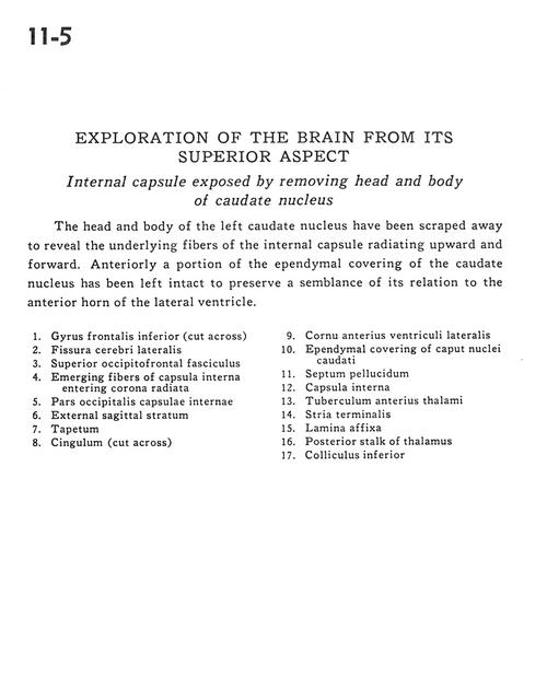
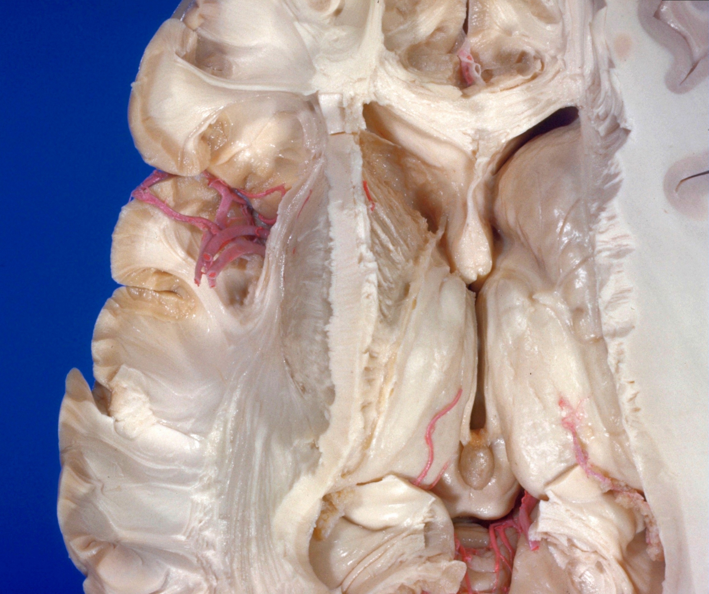
Exploration of the brain from its superior aspect
Internal capsule exposed by removing head and body of caudate nucleus
The head and body of the left caudate nucleus have been scraped away to reveal the underlying fibers of the internal capsule radiating upward and forward. Anteriorly a portion of the ependymal covering of the caudate nucleus has been left intact to preserve a semblance of its relation to the anterior horn of the lateral ventricle.
- Inferior frontal gyrus (cut across)
- Lateral cerebral fissure
- Superior occipitofrontal fasciculus
- Emerging fibers of internal capsule entering corona radiata
- Occipital part internal capsule
- External sagittal stratum
- Tapetum
- Cingulum (cut across)
- Anterior horn lateral ventricle
- Ependymal covering of head of caudate
- Septum pellucidum
- Internal capsule
- Anterior tubercle of thalamus
- Stria terminalis
- Lamina affixa
- Posterior stalk of thalamus
- Inferior colliculus