Dorsal aspect of hand
Dorsal interosseous muscles
Stanford holds the copyright to the David L. Bassett anatomical images and has assigned
Creative Commons license Attribution-Share
Alike 4.0 International to all of the images.
For additional information regarding use and permissions,
please contact Dr. Drew Bourn at dbourn@stanford.edu.
Image #109-5
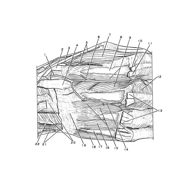
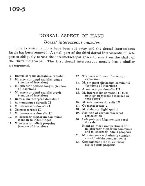
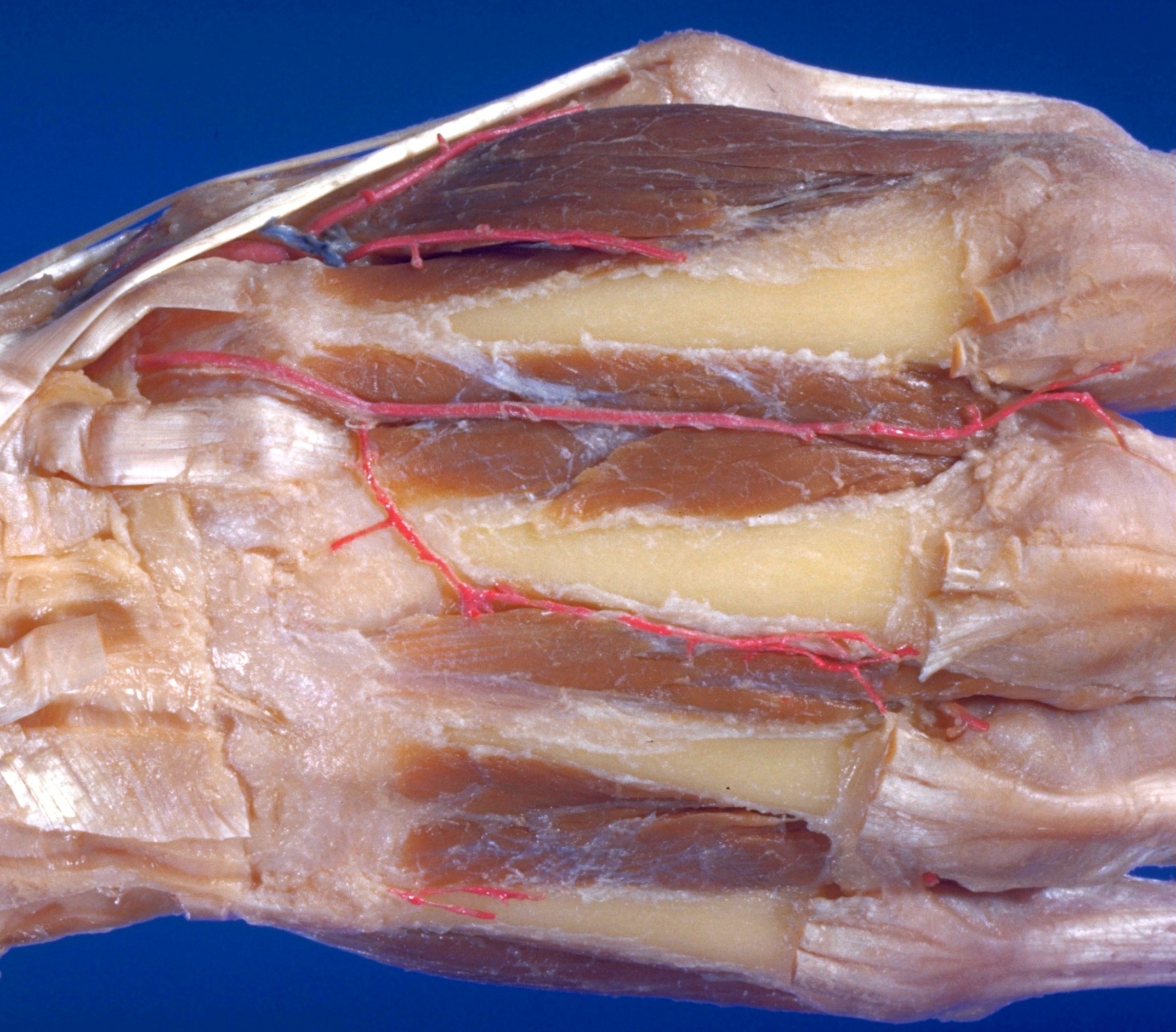
Dorsal aspect of hand
Dorsal interosseous muscles
The extensor tendons have been cut away and the dorsal interosseous fascia has been removed. A small part of the third dorsal interosseous muscle passes obliquely across the intermetacarpal space to insert on the shaft of the third metacarpal. The first dorsal interosseous muscle has a similar arrangement.
- Dorsal carpal branch radial artery
- Extensor carpi radialis longus muscle (tendon of insertion)
- Extensor pollicis longus muscle (tendon of insertion)
- Extensor carpi radialis brevis muscle (tendon of insertion)
- Branches of dorsal metacarpal artery I
- Dorsal metacarpal artery II
- Dorsal interosseous muscle I
- Metacarpal II
- Dorsal interosseous muscle II
- Common extensor digitorum muscle (tendon to index finger)
- Extensor indicis muscle (tendon of insertion)
- Transverse fibers of extensor expansion
- Common extensor digitorum muscle (tendons of insertion)
- Dorsal metacarpal artery III
- Dorsal interosseous muscle III (left pointer on muscle described in text above)
- Dorsal interosseous muscle IV
- Metacarpal V
- Abductor digiti minimi muscle
- Position of carpometacarpal articulation
- Left pointer: Dorsal carpal ligament Right pointer: Compartment for common extensor digitorum muscle and extensor indicis muscle
- Extensor carpi ulnaris muscle (tendon cut off within compartment)
- Compartment for extensor digiti minimi muscle