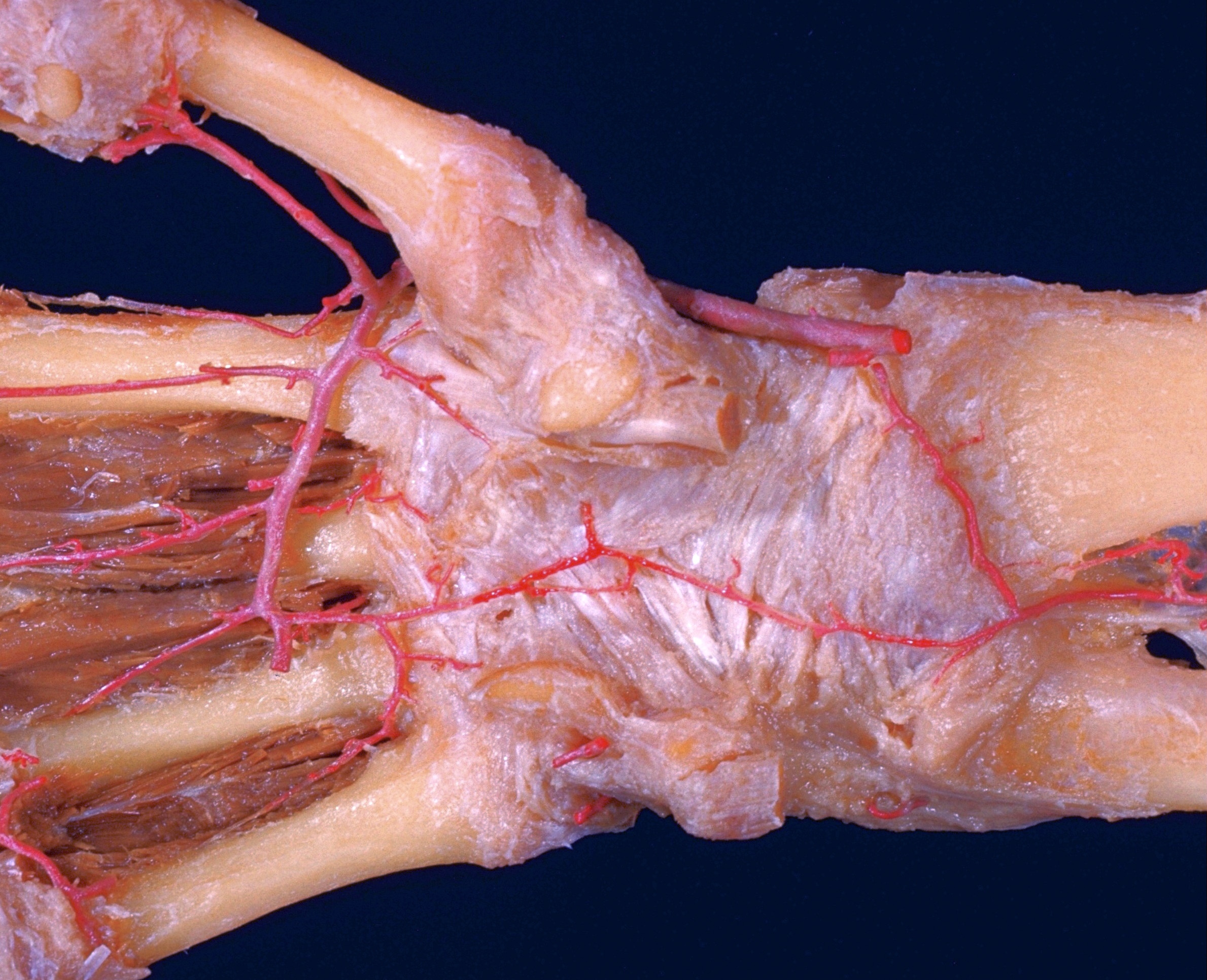
Volar aspect of right hand
Branches of deep volar arch
Stanford holds the copyright to the David L. Bassett anatomical images and has assigned
Creative Commons license Attribution-Share
Alike 4.0 International to all of the images.
For additional information regarding use and permissions,
please contact Dr. Drew Bourn at dbourn@stanford.edu.
Image #105-3
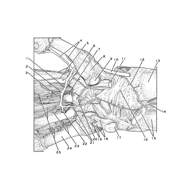
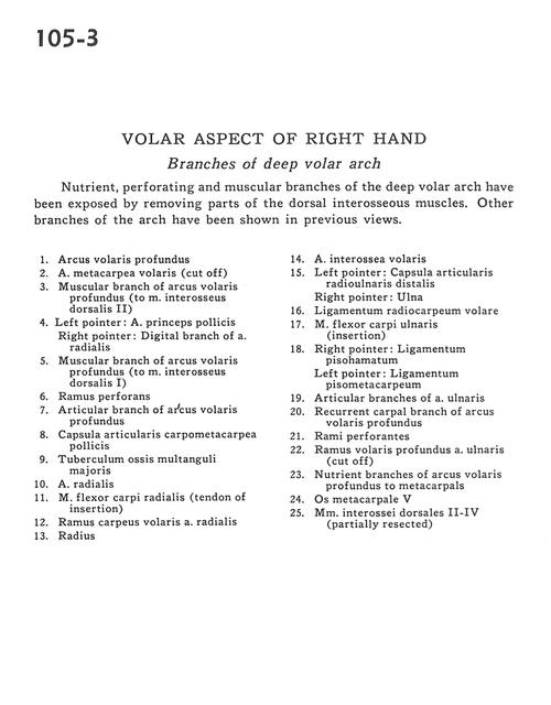
Volar aspect of right hand
Branches of deep volar arch
Nutrient, perforating and muscular branches of the deep volar arch have been exposed by removing parts of the dorsal interosseous muscles. Other branches of the arch have been shown in previous views.
- Deep palmar arch
- Anterior metacarpal artery (cutoff)
- Muscular branch of deep palmar arch (to dorsal interosseous muscle II)
- Left pointer: Princeps pollicis artery Right pointer: Digital branch of radial artery
- Muscular branch of deep palmar arch (to dorsal interosseus muscle I)
- Perforating branch
- Articular branch of deep palmar arch
- Metacarpotrapezial joint capsule
- Tubercle of trapezium bone
- Radial artery
- Flexor carpi radialis muscle (tendon of insertion)
- Anterior carpal branch radial artery
- Radius
- Anterior interosseous artery
- Left pointer: Distal radioulnar joint capsule Right pointer: Ulna
- Anterior radiocarpal ligament
- Flexor carpi ulnaris muscle (insertion)
- Right pointer: Pisohamate ligament Left pointer: Pisometacarpal ligament
- Articular branches of ulnar artery
- Recurrent carpal branch of deep palmar arch
- Perforating branches
- Deep anterior branch ulnar artery (cut off)
- Nutrient branches of deep palmar arch to metacarpals
- Metacarpal V
- Dorsal interosseous muscles II-IV (partially resected)