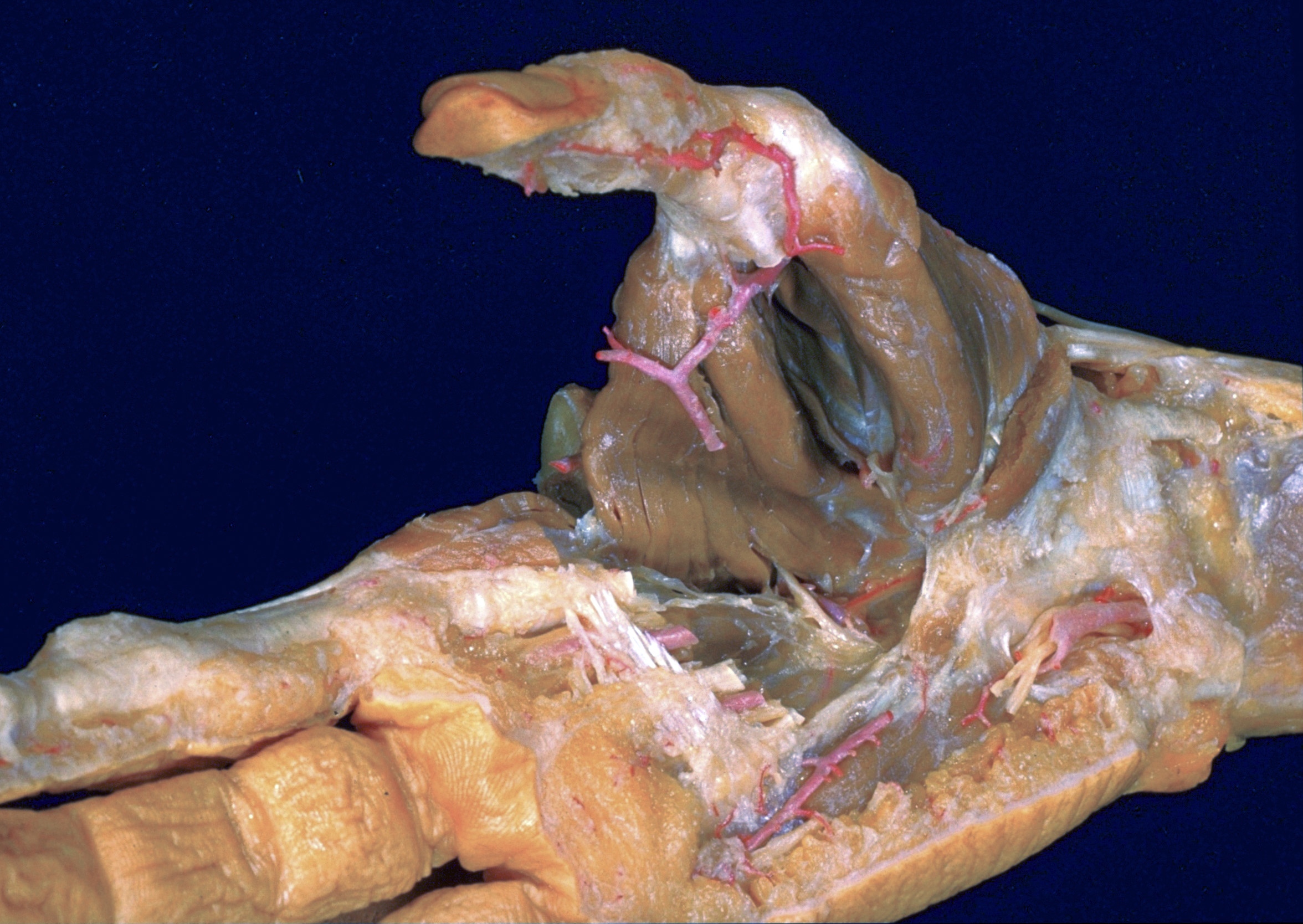
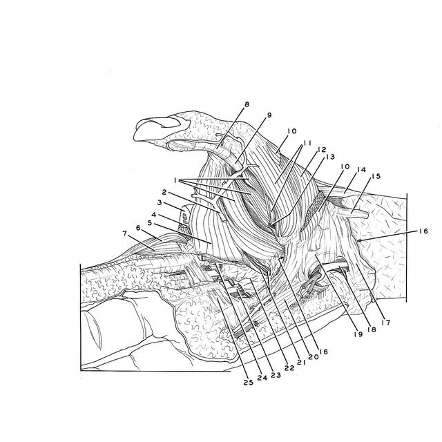
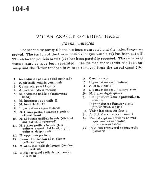
Volar aspect of right hand
Thenar muscles
Stanford holds the copyright to the David L. Bassett anatomical images and has assigned
Creative Commons license Attribution-Share
Alike 4.0 International to all of the images.
For additional information regarding use and permissions,
please contact Dr. Drew Bourn at dbourn@stanford.edu.
Image #104-4
Volar aspect of right hand
Thenar muscles
The second metacarpal bone has been transected and the index finger removed. The tendon of the flexor pollicis longus muscle (9) has been cut off. The abductor pollicis brevis (10) has been partially resected. The remaining thenar muscles have been separated. The palmar aponeurosis has been cut away and the flexor tendons have been removed from the carpal canal (16).
- Adductor pollicis muscle (oblique head)
- Anterior common digital artery
- Metacarpal II (cut)
- Anterior radial index artery
- Adductor pollicis muscle (transverse head)
- Dorsal interosseous muscle
- Lumbrical muscle II
- Ligament of digital sheath
- Flexor pollicis longus muscle (tendon of insertion)
- Abductor pollicis brevis muscle (divided and partially removed)
- Flexor pollicis brevis muscle (left pointer superficial head; right pointer deep head)
- Opponens pollicis muscle
- Groove for tendon of flexor pollicis longus muscle
- Abductor pollicis longus muscle (tendon of insertion)
- Flexor carpi radialis muscle (tendon of insertion)
- Carpal tunnel
- Anterior carpal ligament
- Ulnar artery and nerve
- Transverse carpal ligament
- Flexor digiti minimi muscle
- Left pointer: Deep branch of ulnar nerve Right pointer: Deep anterior branch ulnar artery
- Anterior interosseous fascia
- Anterior common digital artery
- Fascial septum between palmar aponeurosis and anterior interosseous fascia
- Transverse fascicle of palmar aponeurosis