Volar aspect of right hand
Carpal canal opened; fascia intact
Stanford holds the copyright to the David L. Bassett anatomical images and has assigned
Creative Commons license Attribution-Share
Alike 4.0 International to all of the images.
For additional information regarding use and permissions,
please contact Dr. Drew Bourn at dbourn@stanford.edu.
Image #102-7
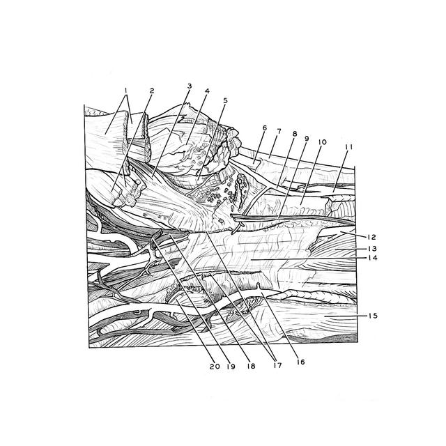
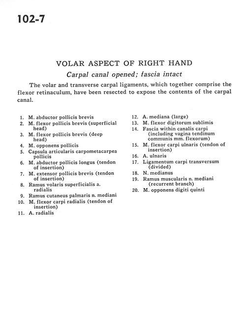
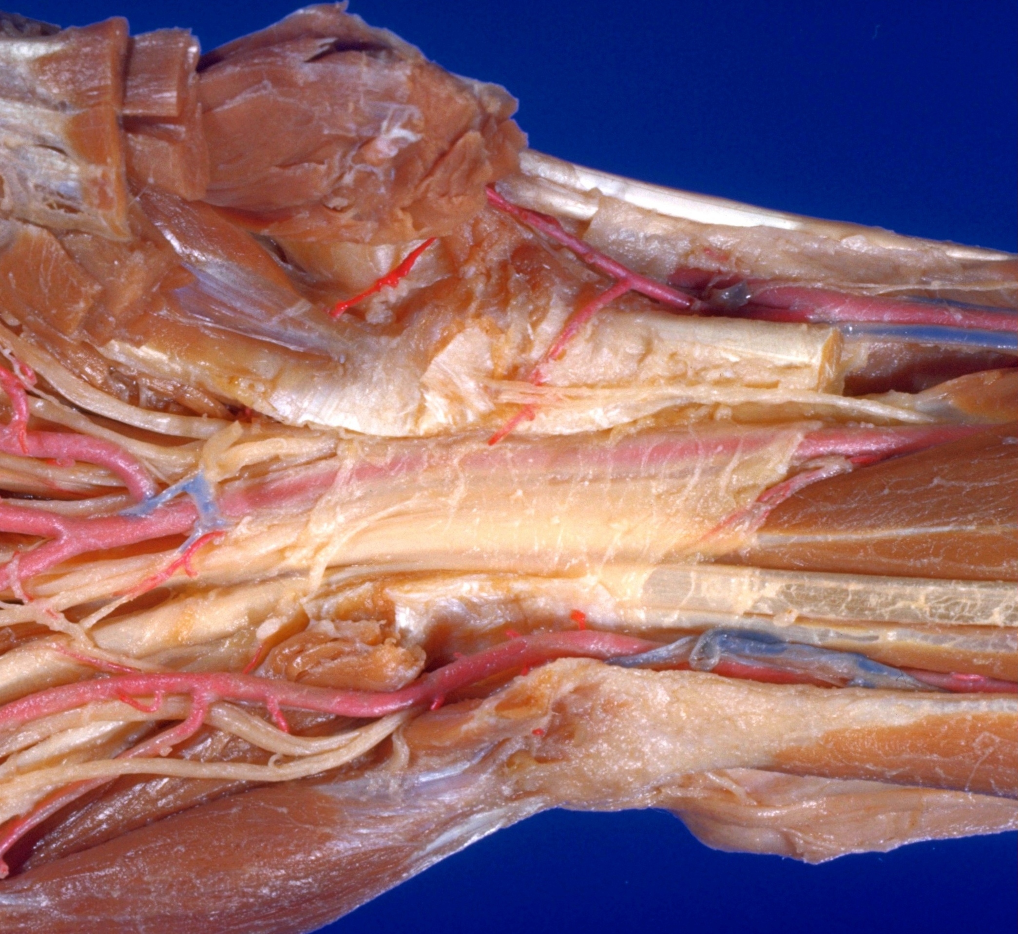
Volar aspect of right hand
Carpal canal opened; fascia intact
The volar and transverse carpal ligaments, which together comprise the flexor retinaculum, have been resected to expose the contents of the carpal canal.
- Abductor pollicis brevis muscle
- Flexor pollicis brevis muscle (superficial head)
- Flexor pollicis brevis muscle (deep head)
- Opponens pollicis muscle
- Metacarpotrapezial joint capsule
- Abductor pollicis longus muscle (tendon of insertion)
- Extensor pollicis brevis muscle (tendon of insertion)
- Superficial anterior branch radial artery
- Palmar cutaneous branch of median nerve
- Flexor carpi radialis muscle (tendon of insertion)
- Radial artery
- Median artery (large)
- Flexor digitorum superficialis muscle
- Fascia within carpal tunnel (including sheath of common tendon of flexor muscles)
- Flexor carpi ulnaris muscle (tendon of insertion)
- Ulnar artery
- Transverse carpal ligament (divided)
- Median nerve
- Muscular branch of median nerve (recurrent branch)
- Opponens digiti minimi muscle