Volar aspect of right hand
Tendon sheath of flexor pollicis longus muscle; branches of ulnar nerve to flexor pollicis brevis muscle
Stanford holds the copyright to the David L. Bassett anatomical images and has assigned
Creative Commons license Attribution-Share
Alike 4.0 International to all of the images.
For additional information regarding use and permissions,
please contact Dr. Drew Bourn at dbourn@stanford.edu.
Image #102-5

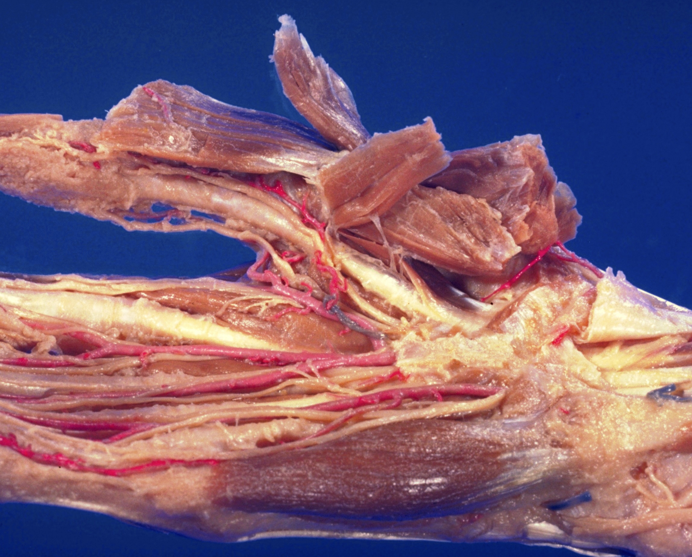
Volar aspect of right hand
Tendon sheath of flexor pollicis longus muscle; branches of ulnar nerve to flexor pollicis brevis muscle
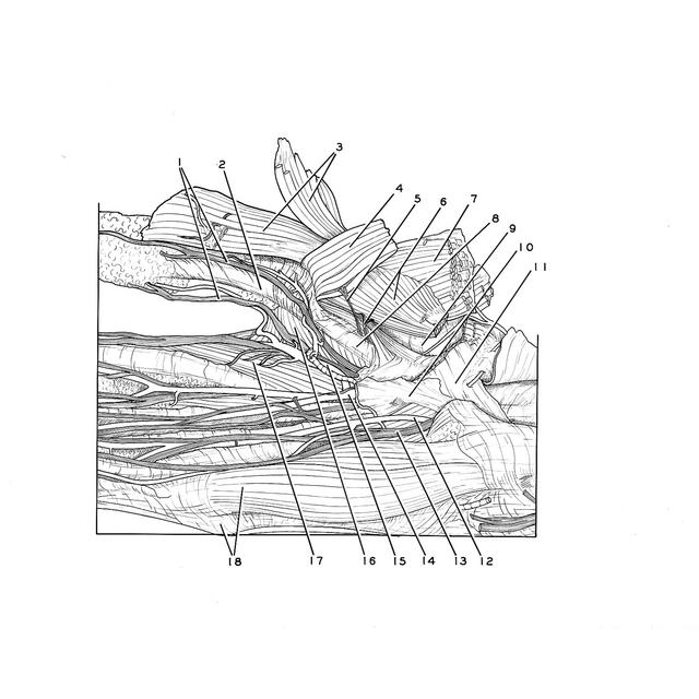
The specimen has been turned to show the medial side of the thumb. The abductor pollicis brevis (3), flexor pollicis brevis (4,6) and opponens pollicis (7) muscles have been reflected in various ways. Air has been injected into the synovial sheath (8) of the flexor pollicis longus.
- Proper palmar digital nerves of median nerve
- Ligament of digital sheath I
- Abductor pollicis brevis muscle (reflected)
- Flexor pollicis brevis muscle (portion of muscle which inserts with abductor pollicis brevis muscle)
- Muscular branch of ulnar nerve (to flexor pollicis brevis muscle)
- Left pointer: Muscular branch of ulnar nerve (to flexor pollicis brevis muscle) Right pointer: Flexor pollicis brevis muscle (superficial head)
- Opponens pollicis muscle
- Tendon sheath of flexor pollicis longus muscle
- Metacarpotrapezial joint capsule
- Palmar aponeurosis
- Transverse carpal ligament
- Ulnar artery
- Ulnar nerve
- Median artery (large)
- Anterior common digital artery (to thumb and index finger)
- Adductor pollicis muscle
- Lumbrical muscle I
- Left pointer: Position of metacarpophalangeal joint of fifth finger Right pointer: Abductor digiti minimi muscle