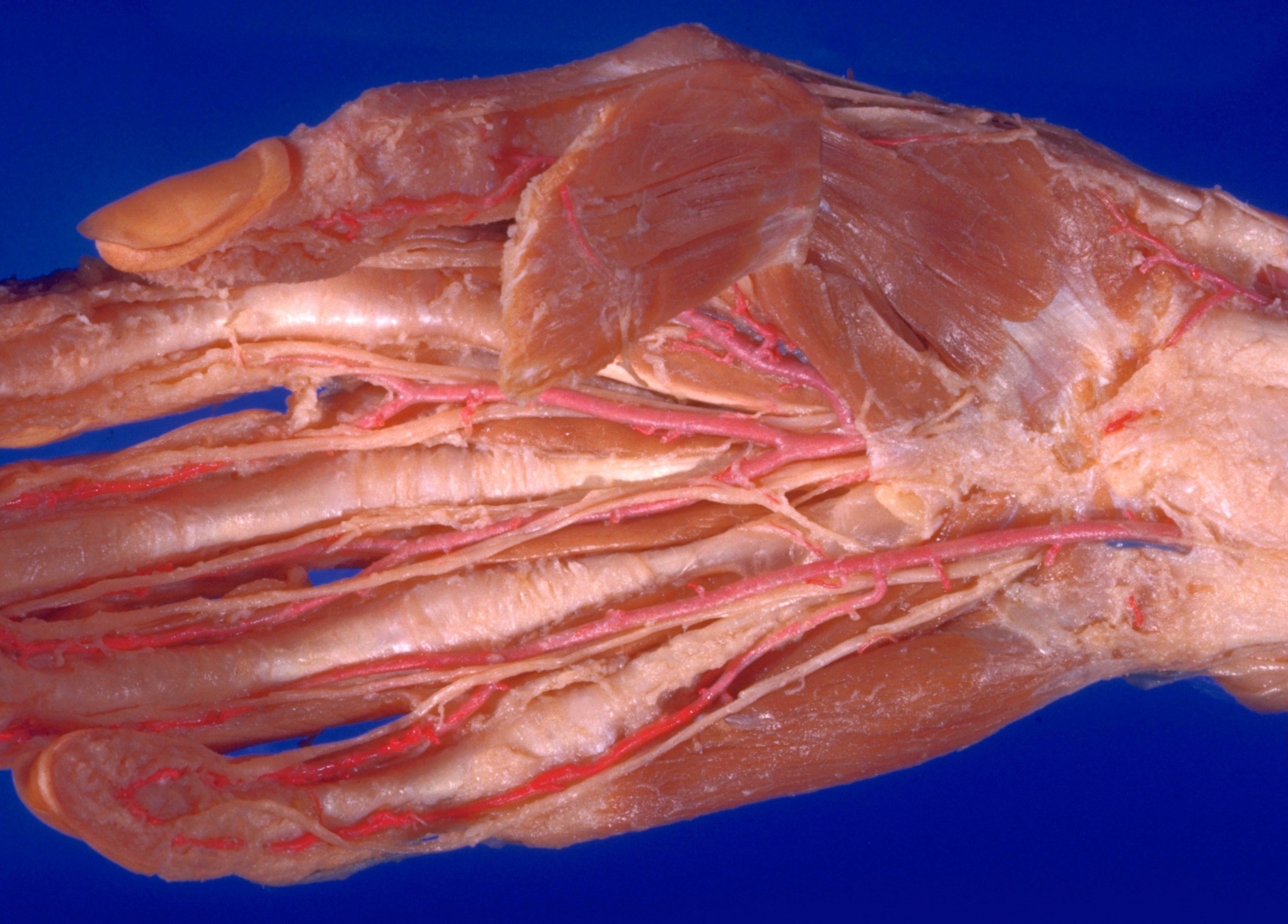
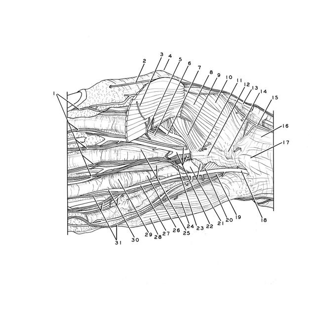
Volar aspect of right hand
Thenar and hypothenar muscles, general view
Stanford holds the copyright to the David L. Bassett anatomical images and has assigned
Creative Commons license Attribution-Share
Alike 4.0 International to all of the images.
For additional information regarding use and permissions,
please contact Dr. Drew Bourn at dbourn@stanford.edu.
Image #102-2
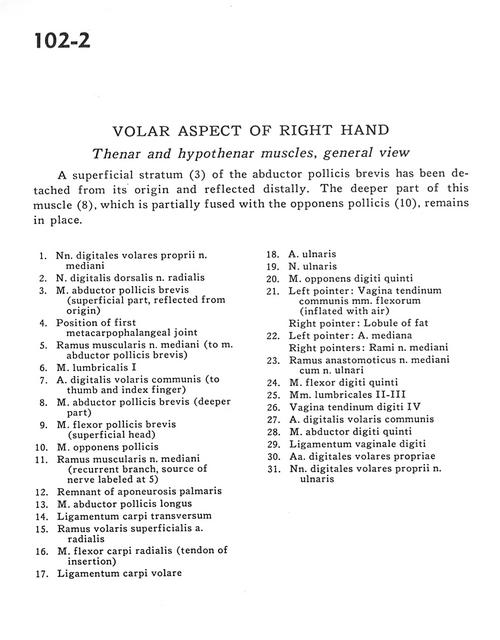
Volar aspect of right hand
Thenar and hypothenar muscles, general view
A superficial stratum (3) of the abductor pollicis brevis has been detached from its origin and reflected distally. The deeper part of this muscle (8), which is partially fused with the opponens pollicis (10), remains in place.
- Proper palmar digital nerves of median nerve
- Dorsal digital nerve of radial nerve
- Abductor pollicis brevis muscle (superficial part, reflected from origin)
- Position of first metacarpophalangeal joint
- Muscular branch of median nerve (to abductor pollicis brevis muscle)
- Lumbrical muscle
- Anterior common digital artery (to thumb and index finger)
- Abductor pollicis brevis muscle (deeper part)
- Flexor pollicis brevis muscle (superficial head)
- Opponens pollicis muscle
- Muscular branch of median nerve (recurrent branch source of nerve labeled at 5)
- Remnant of palmar aponeurosis
- Abductor pollicis longus muscle
- Transverse carpal ligament
- Superficial anterior branch radial artery
- Flexor carpi radialis muscle (tendon of insertion)
- Anterior carpal ligament
- Ulnar artery
- Ulnar nerve
- Opponens digiti minimi muscle
- Left pointer: Sheath of common tendon of flexor muscles (inflated with air) Right pointer: Lobule of fat
- Left pointer: Median artery Right pointers: Branches of median nerve
- Anastomotic branch of median nerve with ulnar nerve
- Flexor digiti minimi muscle
- Lumbrical muscles II-III
- Tendon of digital sheath IV
- Anterior common digital artery
- Abductor digiti minimi muscle
- Ligament of digital sheath
- Anterior proper digital arteries
- Proper palmar digital nerves of ulnar nerve